©2010 Baishideng.
World J Gastrointest Pathophysiol. Jun 15, 2010; 1(2): 50-62
Published online Jun 15, 2010. doi: 10.4291/wjgp.v1.i2.50
Published online Jun 15, 2010. doi: 10.4291/wjgp.v1.i2.50
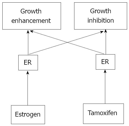
Figure 1 Two-pathway theory of estrogen-receptor-dependent growth.
Many estrogen target cells contain growth-stimulatory and -inhibitory pathways. Both pathways are mediated through ERs. Tamoxifen may possess relatively high affinity for the growth-inhibitory pathway whereas estrogen can mainly activate the growth-stimulatory pathway. However, any ligand for ER can stimulate both pathways.
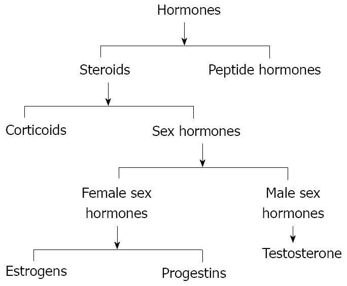
Figure 2 Scheme of the two classes of hormones, steroids and peptide hormones that successively can be divided in corticoids and sex hormones.
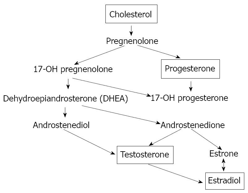
Figure 3 The biosynthesis of the sex hormones starts with the oxidation of the side chain of cholesterol, which is catalyzed by the enzyme cytochrome P450scc to form pregnenolone.
The next steps in the biosynthesis of testosterone can proceed via two different routes. Pregnenolone can be oxidised first by cytochrome P45017a to 17a-hydroxypregnenolone. The enzyme 3β-HSD also can convert pregnenolone first into progesterone. Both pregnenolone and progesterone are accepted as substrate by the enzyme cytochrome P45017a. In this way, after 3β-hydroxy-5-androstene-17-one (DHEA) synthesis, there is the testosterone and successively the estradiol formation.
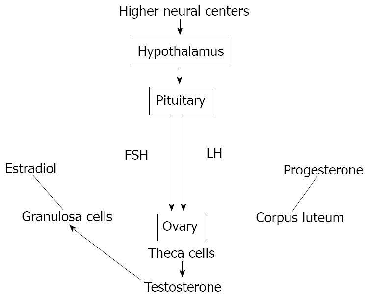
Figure 4 Scheme of hypothalamic-pituitary-gonadal (HPG) axis control exerted by both circulating and in situ locally produced estradiol in men.
This axis controls development, reproduction, and aging in animals. The hypothalamus produces gonadotropin-releasing hormone (GnRH). The pituitary gland produces luteinizing hormone (LH) and follicle-stimulating hormone (FSH), and the gonads produce estrogen, progesterone and testosterone from different kinds of cells.
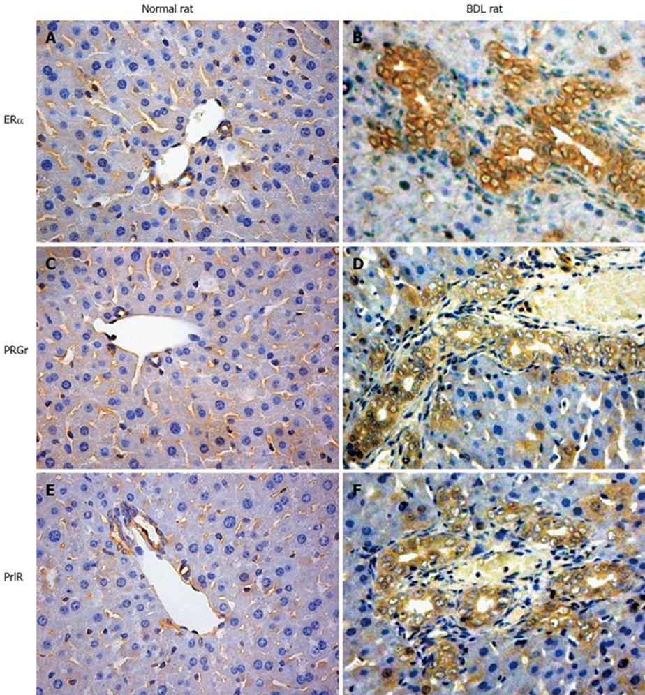
Figure 5 Some representative immunohistochemistry for Erα, progesterone and prolactin receptors in normal and bile duct ligation (BDL) rats.
A, B: Erα receptors; C, D: progesterone receptors; E, F: prolactin receptors. The expression of all these receptors is highly increased after BDL compared with that in the biliary epithelium of the normal animal. Original magnification × 40.
Figure 6 Immunohistochemistry for ERα, progesterone and prolactin receptors in liver sections from patients affected with polycystic liver disease.
A: Erα receptors; B: progesterone recepors; C: prolactin receptors. Also in course of human cholangiopathies, these three considered receptors seem to play an important role in cholangiocyte physiology. Original magnification × 20.
- Citation: Mancinelli R, Onori P, DeMorrow S, Francis H, Glaser S, Franchitto A, Carpino G, Alpini G, Gaudio E. Role of sex hormones in the modulation of cholangiocyte function. World J Gastrointest Pathophysiol 2010; 1(2): 50-62
- URL: https://www.wjgnet.com/2150-5330/full/v1/i2/50.htm
- DOI: https://dx.doi.org/10.4291/wjgp.v1.i2.50


