Published online Jul 28, 2017. doi: 10.4329/wjr.v9.i7.304
Peer-review started: December 31, 2016
First decision: March 28, 2017
Revised: April 16, 2017
Accepted: May 30, 2017
Article in press: June 1, 2017
Published online: July 28, 2017
Processing time: 205 Days and 5.2 Hours
To validate the feasibility of high resolution computed tomography (HRCT) of the lung prior to computed tomography angiography (CTA) in assessing incidental thoracic findings during endovascular aortic aneurysm repair (EVAR) planning or follow-up.
We conducted a retrospective study among 181 patients (143 men, mean age 71 years, range 50-94) referred to our centre for CTA EVAR planning or follow-up. HRCT and CTA were performed before or after 1 or 12 mo respectively to EVAR in all patients. All HRCT examinations were reviewed by two radiologists with 15 and 8 years’ experience in thoracic imaging. The results were compared with histology, bronchoscopy or follow-up HRCT in 12, 8 and 82 nodules respectively.
There were a total of 102 suspected nodules in 92 HRCT examinations, with a mean of 1.79 nodules per patient and an average diameter of 9.2 mm (range 4-56 mm). Eighty-nine out of 181 HRCTs resulted negative for the presence of suspected nodules with a mean smoking history of 10 pack-years (p-y, range 5-18 p-y). Eighty-two out of 102 (76.4%) of the nodules met criteria for computed tomography follow-up, to exclude the malignant evolution. Of the remaining 20 nodules, 10 out of 20 (50%) nodules, suspected for malignancy, underwent biopsy and then surgical intervention that confirmed the neoplastic nature: 4 (20%) adenocarcinomas, 4 (20%) squamous cell carcinomas, 1 (5%) small cell lung cancer and 1 (5%) breast cancer metastasis); 8 out of 20 (40%) underwent bronchoscopy (8 pneumonia) and 2 out of 20 (10%) underwent biopsy with the diagnosis of sarcoidosis.
HRCT in EVAR planning and follow-up allows to correctly identify patients requiring additional treatments, especially in case of lung cancer.
Core tip: Nowadays the use of high resolution computed tomography in endovascular aortic aneurysm repair (EVAR) patients planning and follow up is not recommended yet. Our study demonstrates the possibility to early diagnose lung cancer during EVAR follow-up or planning in smoker patients, overcoming the concept of dose radiation induced neoplasms, especially in over 65 years old patients.
- Citation: Mazzei MA, Guerrini S, Gentili F, Galzerano G, Setacci F, Benevento D, Mazzei FG, Volterrani L, Setacci C. Incidental extravascular findings in computed tomographic angiography for planning or monitoring endovascular aortic aneurysm repair: Smoker patients, increased lung cancer prevalence? World J Radiol 2017; 9(7): 304-311
- URL: https://www.wjgnet.com/1949-8470/full/v9/i7/304.htm
- DOI: https://dx.doi.org/10.4329/wjr.v9.i7.304
Vascular diseases cover an extended selection of pathologies comprising cardiovascular, thoracoabdominal, peripheral vascular and cerebrovascular disease[1]. Smoking is considered one of the main risk factors for the development of atherosclerosis and, in particular, oxidative stress and inflammation that constitute the physiological connection between smoking and vascular diseases. Polycyclic aromatic hydrocarbons (PAH) represent the main carcinogenic compound found in cigarettes, produced during the incomplete combustion of organic matter. Many articles demonstrate the double activity of PAH, both carcinogenic (lung and other tissues) and inflammatory, provoking endothelial dysfunction and several studies demonstrate that oxidants directly impair endothelial function, increasing nitric oxide scavenging by oxygen free radicals[2-6]. In the literature it is well known that cardiovascular diseases are often diagnosed on the basis of imaging findings, such as suspected atherosclerotic plaques of the chest in computed tomography (CT), also incidentally[7-9]. A similar approach should be used to identify extravascular findings, when the CT examination is required to explore vascular diseases. In particular, since cigarette smoking is the main risk factor for both vascular and neoplastic lung diseases, radiologists should examine the chest in evaluation of vascular patients with many risk factors suggesting possible synchronous pathologies. It has been reported that CTs requested for the exclusion of pulmonary embolism give a high yield of chest abnormalities, such as mediastinal adenopathy, paratracheal adenopathy, atelectasis, emphysema and pulmonary nodules or masses[10,11]. Although not the target of the investigation, lung abnormalities, especially lung cancer, could become the main finding with prognostic relevance in terms of life-long survivor risk in vascular patients. Furthermore, even if many articles over the last decade have reported the problem of unsuspected thoracic findings in CTs performed for suspected pulmonary embolism or thoracic aortic pathology, the possibility of finding chest pathology, and in particular lung cancer in smoker patients suffering from vascular disease, has been underestimated[12,13]. Moreover, several articles have reported a high cumulative radiation dose in patients treated with endovascular aneurysm repair (EVAR), both during the interventional procedure and CT follow-up, suggesting a possible role of radiation exposure in developing cancer in these patients[14-16]. On the contrary, there have been no articles to date about the discovery of lung cancer during EVAR planning or surveillance in smokers suggesting that smoking rather than the exposure of patients to radiation is the main risk factor for lung cancer. Considering the previous statements, the purpose of this study is to assess the prevalence of lung cancer in patients with a smoking attitude, who underwent computed tomography angiography (CTA) for planning or monitoring EVAR in our department.
Institutional review board approval was obtained for this retrospective study, as well as informed consent from all subjects. We reviewed the report results of 250 CTs of patients referred to our department for EVAR planning or follow-up between June 2014 and May 2016, searching for lung abnormalities in the patients who underwent high resolution computed tomography (HRCT) of the lung before contrast agent administration. Patients were identified throughout a digital radiological database (Picture Archive and Communication System, PACS) which registers all radiological studies performed by the Department of Radiology. The mean age of patients at the time of CT was 71 years, in a range from 50 to 94, and 38 (21%) were female. Sixty-nine (26.6%) patients were excluded for the following reasons: 30 (43.5%) because of the absence of synchronous chest HRCT, 21 (30.4%) because of lack of proven histological findings of lung cancer or HRCT follow-up examinations, and 18 (26.1%) because of non-smoking attitude. The CTA and HRCT were performed simultaneously in all selected cases to avoid interpretation bias. Seventy-four (40.8%) CTs were performed for EVAR planning and 107 for EVAR surveillance (67 at 1 mo and 40 at 12 mo after EVAR). All the selected patients had a documented history of smoking [number of pack-years (p-y)][17-19].
All the CTs were performed with a 64-detector row CT scanner (Discovery HD 750, General Electric Healthcare, and Milwaukee, United States). HRCTs were acquired at end of inspiration using volumetric technique in the caudocranial direction from the basis to the apex of the lung; patients were in supine position. The following technical parameters were used: Effective slice thickness 3.75 mm, collimation 40 mm, beam pitch 0.969, reconstruction interval 1 mm, tube voltage 140 kVp and reference mAs 250/400. Automatic tube current modulation was used to minimise radiation exposure. Chest CTs were acquired using a standard algorithm, then data were reconstructed by using a high spatial-frequency algorithm (bone plus), with 1.25 mm slice thickness. Abdominal CT angiography (CTA) was performed with a spiral technique in the caudocranial direction (from the pelvic brim to the lung bases) with the patients supine. Patients were instructed to hold their breath during helical imaging to avoid motion artefacts. After a scout-view scan, intravenous injection of 1.5 mL/kg non-ionic contrast material (Iomeprol 400 mg iodine/mL; Iomeron 400, Bracco Diagnostics, Milan, Italy) followed by 40 mL saline solution was administered with an 18-gauge needle in the antecubital vein, using a dual-barrel injector (4 mL/s flow rate, CT Motion, Ulrich Medical, Ulm, Germany). Arterial phase images were obtained 4 seconds after bolus detection in the suprarenal aorta. The following technical parameters were used: Effective slice thickness 1.25 mm, collimation 40 mm, beam pitch 0.969, reconstruction interval 0.8 mm, tube voltage 140 kVp and reference mAs 250/700. A standard reconstruction algorithm was used. In 24 out of 181 patients (13.2%), the contrast CT was extended to the thorax using the same technical parameters. Automatic tube current modulation was also used to minimise radiation exposure in the post-contrast examination[16].
All images were analysed independently and blindly by two readers with 15 and 8 years’ experience in chest-imaging respectively. HRCT scans were analysed on a reconstruction and image interpretation console (Advantage Workstation 4.4, GE Healthcare, Milwaukee, Wis, United States), adjusting the image level, window and enlargement values each time, and routinely using a 2D multiplanar reconstruction technique (coronal, sagittal and oblique planes). Pulmonary HRCT findings included: Pleural effusion, atelectasis/pneumonia, pericardial effusion, cardiomegaly, coronary artery calcifications, bone findings, hiatal hernia, emphysema, mediastinal or hilar adenopathy, pulmonary micronodule (< 4 mm), pulmonary nodule (> 4 mm and < 30 mm) and pulmonary mass (> 30 mm). The readers recorded any incidental finding, with particular attention to pulmonary nodules or masses and differences were resolved by consensus. All nodules were characterised by number, size (measured in their greatest diameter) and CT characteristic appearance (solid, ground-glass or partially solid, edge characteristics, speculated or smooth, presence or absence of pleural-tag, bronchus sign, calcifications, intralesional fat or intralesional air) and reviewed with the smoking history of the patient. According to Fleischner Society guidelines, all suspected nodules were addressed to CT biopsy or surgical intervention, whereas nodules with a CT low risk appearance for lung cancer were addressed to CT follow-up[20] (Table 1, Table 2, Table 3, Table 4). All previous medical reports, clinical notes, discharges summaries and medical histories of patients were examined to potentially define every mass or nodule as a new incidental finding.
| Nodule size (mm) | Ground-glass | Part solid |
| < 6 | No routine FU | No routine FU |
| ≥ 6 | CT at 6-12 mo to confirm persistence, then CT every 2 yr until 5 yr | CT at 3-6 mo to confirm persistence. If unchanged and solid component remains <6 mm, annual CT should be performed for 5 yr |
| Nodule size (mm) | |
| < 6 | CT at 3-6 mo. If stable, consider CT at 2 and 4 yr |
| ≥ 6 | CT at 3-6 mo. Subsequent management based on the most suspicious nodule(s) |
The lung findings detected by the readers were collected, and the results expressed as mean ± SD. A descriptive statistical analysis was performed; the quantitative variables were expressed as means and range whereas the qualitative values as percentages. The statistical review of the study was performed by a biomedical statistician. The analysis was performed using Stata version 8.0 (Stata Corp, College Station, Texas).
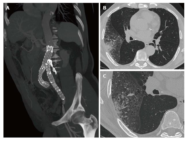
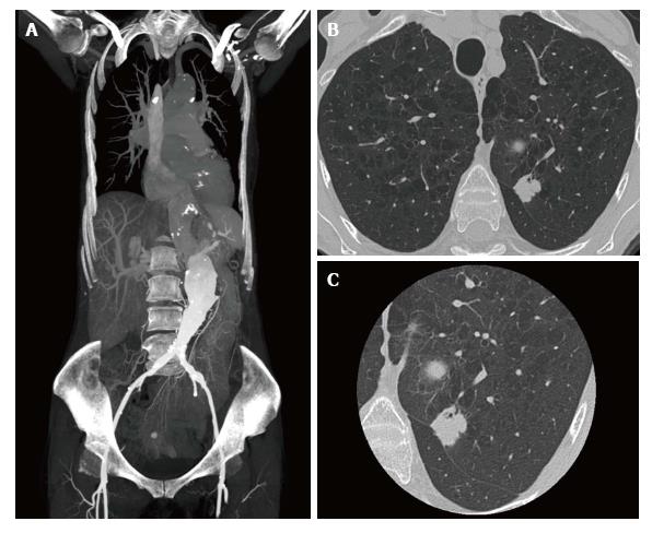
A total of 181 HRCTs were reviewed. The incidental lung findings reported on chest CT are shown in Table 5. There were a total of 102 (56%) nodules in 92 out of 181 (50.8%) HRCTs, with a mean of 1.79 nodules per patient and an average diameter of 9.2 mm, ranging from 4 to 56 mm. All the CT nodules characteristics are reported in Table 6. After radiologists’ review, 82 (76.4%) of the nodules met criteria for CT follow-up and were submitted to a second HRCT examination (performed between 6-12 mo), to exclude the possibility of malignant evolution. Of the remaining 20 nodules, 10 out of the 20 (50%) suspected for malignancy underwent biopsy and then surgical intervention which confirmed the following neoplastic nature: 4 (20%) adenocarcinomas (Figure 1), 4 (20%) squamous cell carcinomas, 1 (5%) small cell lung cancer and 1 (5%) breast cancer metastasis (Figure 2); 8 out of 20 (40%) underwent bronchoscopy (8 pneumonia) and 2 out of 20 (10%) underwent biopsy with the diagnosis of sarcoidosis. All the patients diagnosed with lung cancer (1 female and 8 males) had a smoking history with a mean quantity of 60 p-y (range 45-83 p-y). The remaining patients with non-neoplastic nodules had a smoking history with a mean quantity of 35 p-y (range 20-46 p-y) per patient. Eighty-nine out of 181 HRCTs resulted negative for the presence of suspected nodules with a mean smoking history of 10 p-y (range 5-18 p-y).
| Patients (n) | Findings |
| 31 | Pleural effusion |
| 5 | Atelectasis |
| 8 | Pneumonia |
| 16 | Pericardial effusion |
| 48 | Cardiomegaly |
| 57 | Coronary artery calcifications |
| 0 | Bone findings |
| 23 | Hiatal hernia |
| 94 | Emphysema |
| 39 | Mediastinal or hilar adenopathy |
| Nodules size | n (tot 102) |
| Pulmonary micronodule (< 4 mm) | 43 |
| Pulmonary nodule (> 4 mm and < 30 mm) | 51 |
| Pulmonary mass (> 30 mm) | 8 |
| Nodules characteristics | n |
| Solid | 73 |
| Ground-glass | 21 |
| Partially solid | 8 |
| Spiculated | 9 |
| Smooth | 4 |
| Pleural tag | 3 |
| Bronchus sign | 6 |
| Calcifications | 7 |
| Intralesion fat | 4 |
| Intralesional air | 2 |
EVAR currently represents a safe and effective treatment for abdominal aortic aneurysms exclusion with an increase in the choice of this treatment over traditional open repair, especially in elderly patients[21,22]. In particular, EVAR is also becoming the method of choice for aneurysmal sac exclusion in vascular patients with difficult vascular anatomies due to its favourable outcomes, customised approach and easy technical execution[23-25]. Despite these advantages some articles debate the risk of long-term lifelong EVAR CT follow-up, with a remarkable amount of radiation exposure carrying the risk of developing cancers; moreover they report the need of dose optimisations using new targeted CT protocols, considering that the absorbed dose by the patient differs on the basis different scanners, patient body size and age[26-28]. However to our knowledge there are no articles discussing the prevalence of lung cancer, prior to EVAR treatment, in patients with a smoking attitude. Furthermore, lung cancer represents the most important cause of death in the world, and the majority of patients suffering from lung cancer present mild or no symptoms, with nodular lesions being the most common presentation of peripheral lung adenocarcinoma[12]. Moreover, a large number of patients with vascular disease have a smoking history and, in particular, smoking is one of the major risk factors for developing vascular diseases. At the same time, smoking also represents the main risk factor for lung cancer due to the activation of the same inflammatory pathway with continuous endothelial damage. In our study we assess the prevalence of lung nodules through HRCT in a cohort of smoker patients who underwent abdominal CTA for planning or follow-up EVAR; considering the mean advanced age of patients (71 years), stochastic radiation damage deriving from the extra dose of HRCT (respectively mean CTDI 12.6 mGy (range 9.4-15.2) for HRCT vs 22.3 mGy (19.8-24.3) for abdominal CTA) can be considered negligible, comparing to the benefit of early tumor detection; in fact 9 out of 102 nodules (8.8%) in 9 out of 181 (4.9%) patients were finally diagnosed as lung cancer with consequent surgery (6 patients), chemotherapeutic treatment (1 patient) or both (2 patients), with a free survivor rate of 100% at one years. All the other patients were correctly addressed to the appropriate treatment or follow-up. In this context, performing HRCT of the lung to optimise morphological evaluation of lung nodules and/or adding advanced imaging procedure such as CT perfusion or CT volumetric assessment of lung nodules during CTA for the evaluation of vascular aneurysm, offers radiologists the possibility of performing a differential diagnosis between benign or malignant nodules, choosing the correct management for each patient[29-33]. Moreover, CTA can be performed in the emergency setting to exclude or confirm the presence of aneurysmal sac or EVAR complications or to investigate an abdominal pain after a doubtful ultrasound examination; incidental findings can also occur in that setting[34-37].
In fact several articles in the literature have reported the possibility of discovering coexistent neoplasms such as gastrointestinal and gall bladder cancers during CT examination of vascular patients[38-40].
Our study had some limitations. First of all, it is a retrospective study with some possible bias in patient selection, although the database used to find the patients was complete in medical records. Secondly, not all the patients underwent a prior chest radiography that could exclude patients with negative reports from the study. Additionally, a prospective study is mandatory to test the real impact of incidental findings in smoker patients who underwent EVAR planning or follow-up, in order to support the introduction of HRCT of the lung in the CT protocol for smoker patients.
In conclusion, this study actually demonstrates the possibility of early diagnosis of lung cancers during CTA EVAR planning or follow-up, overcoming the concept of dose radiation induced neoplasms, especially in patients over the age of 65.
Vascular diseases include an extended selection of pathologies (cardiovascular disease, thoraco-abdominal, peripheral vascular disease and brain vascular disease) and smoking is considered one of the main risk factor for the development of atherosclerosis and in particular oxidative stress and inflammation that provide the physiological connection between smoking and vascular diseases. Even if many articles reported the problem of unsuspected thoracic findings at computed tomographies performed for suspected pulmonary embolism or thoracic aortic pathology, the possibility to find chest pathology, and in particular lung cancer in smoker patients suffering from vascular disease, is underestimated.
Nowadays the use of high resolution computed tomography (HRCT) in endovascular aortic aneurysm repair (EVAR) patients planning and follow up is not recommended yet but this study demonstrates the possibility to early diagnose lung cancers during EVAR planning or follow-up, overcoming the concept of dose radiation induced neoplasms, especially in over 65 years old patients.
To be known, this is the first study using HRCT for the evaluation of vascular patients during EVAR planning or follow-up.
HRCT could play a key role in the diagnosis of incidental lung findings during the evaluation of vascular patients (EVAR planning or follow-up) and surely in the management. In particular HRCT can discriminate patients with urgent lung surgical evaluation for the presence of malignancy, from patients in which a follow-up can be proposed, increasing lifelong patients expectancy.
HRCT (high resolution computed tomography of the lung) before contrast agent administration, can lead a better evaluation of lung abnormalities enhancing nodule morphology and shape; CTA allows a better evaluation of vascular lumen during surgical planning and a correct assessment in cases of EVAR follow-up complications.
The manuscript is well written.
Manuscript source: Invited manuscript
Specialty type: Radiology, nuclear medicine and medical imaging
Country of origin: Italy
Peer-review report classification
Grade A (Excellent): 0
Grade B (Very good): B
Grade C (Good): C
Grade D (Fair): D
Grade E (Poor): 0
P- Reviewer: Gao BL, Razek AAKA, Tarazov PG S- Editor: Ji FF L- Editor: A E- Editor: Lu YJ
| 1. | Pini A, Boccalini G, Baccari MC, Becatti M, Garella R, Fiorillo C, Calosi L, Bani D, Nistri S. Protection from cigarette smoke-induced vascular injury by recombinant human relaxin-2 (serelaxin). J Cell Mol Med. 2016;20:891-902. [RCA] [PubMed] [DOI] [Full Text] [Full Text (PDF)] [Cited by in Crossref: 30] [Cited by in RCA: 27] [Article Influence: 2.7] [Reference Citation Analysis (0)] |
| 2. | Michael Pittilo R. Cigarette smoking, endothelial injury and cardiovascular disease. Int J Exp Pathol. 2000;81:219-230. [RCA] [PubMed] [DOI] [Full Text] [Cited by in Crossref: 152] [Cited by in RCA: 165] [Article Influence: 6.3] [Reference Citation Analysis (0)] |
| 3. | Burke A, Fitzgerald GA. Oxidative stress and smoking-induced vascular injury. Prog Cardiovasc Dis. 2003;46:79-90. [RCA] [PubMed] [DOI] [Full Text] [Cited by in Crossref: 180] [Cited by in RCA: 179] [Article Influence: 7.8] [Reference Citation Analysis (0)] |
| 4. | Paraskevas KI, Mikhailidis DP, Veith FJ. Patients with peripheral arterial disease, abdominal aortic aneurysms and carotid artery stenosis are at increased risk for developing lung and other cancers. Int Angiol. 2012;31:404-405. [PubMed] |
| 5. | Ambrose JA, Barua RS. The pathophysiology of cigarette smoking and cardiovascular disease: an update. J Am Coll Cardiol. 2004;43:1731-1737. [RCA] [PubMed] [DOI] [Full Text] [Cited by in Crossref: 1527] [Cited by in RCA: 1670] [Article Influence: 75.9] [Reference Citation Analysis (0)] |
| 6. | Messner B, Bernhard D. Smoking and cardiovascular disease: mechanisms of endothelial dysfunction and early atherogenesis. Arterioscler Thromb Vasc Biol. 2014;34:509-515. [RCA] [PubMed] [DOI] [Full Text] [Cited by in Crossref: 540] [Cited by in RCA: 761] [Article Influence: 63.4] [Reference Citation Analysis (0)] |
| 7. | Bruzzi JF, Rémy-Jardin M, Delhaye D, Teisseire A, Khalil C, Rémy J. When, why, and how to examine the heart during thoracic CT: Part 1, basic principles. AJR Am J Roentgenol. 2006;186:324-332. [RCA] [PubMed] [DOI] [Full Text] [Cited by in Crossref: 34] [Cited by in RCA: 30] [Article Influence: 1.5] [Reference Citation Analysis (0)] |
| 8. | Sverzellati N, Arcadi T, Salvolini L, Dore R, Zompatori M, Mereu M, Battista G, Martella I, Toni F, Cardinale L. Under-reporting of cardiovascular findings on chest CT. Radiol Med. 2016;121:190-199. [RCA] [PubMed] [DOI] [Full Text] [Cited by in Crossref: 29] [Cited by in RCA: 38] [Article Influence: 3.5] [Reference Citation Analysis (0)] |
| 9. | Lee SH, Seo JB, Kang JW, Chae EJ, Park SH, Lim TH. Incidental cardiac and pericardial abnormalities on chest CT. J Thorac Imaging. 2008;23:216-226. [RCA] [PubMed] [DOI] [Full Text] [Cited by in Crossref: 19] [Cited by in RCA: 19] [Article Influence: 1.1] [Reference Citation Analysis (0)] |
| 10. | Hall WB, Truitt SG, Scheunemann LP, Shah SA, Rivera MP, Parker LA, Carson SS. The prevalence of clinically relevant incidental findings on chest computed tomographic angiograms ordered to diagnose pulmonary embolism. Arch Intern Med. 2009;169:1961-1965. [RCA] [PubMed] [DOI] [Full Text] [Cited by in Crossref: 164] [Cited by in RCA: 172] [Article Influence: 10.1] [Reference Citation Analysis (0)] |
| 11. | Richman PB, Courtney DM, Friese J, Matthews J, Field A, Petri R, Kline JA. Prevalence and significance of nonthromboembolic findings on chest computed tomography angiography performed to rule out pulmonary embolism: a multicenter study of 1,025 emergency department patients. Acad Emerg Med. 2004;11:642-647. [RCA] [PubMed] [DOI] [Full Text] [Cited by in Crossref: 45] [Cited by in RCA: 37] [Article Influence: 1.7] [Reference Citation Analysis (0)] |
| 12. | Kino A, Boiselle PM, Raptopoulos V, Hatabu H. Lung cancer detected in patients presenting to the Emergency Department studies for suspected pulmonary embolism on computed tomography pulmonary angiography. Eur J Radiol. 2006;58:119-123. [RCA] [PubMed] [DOI] [Full Text] [Cited by in Crossref: 11] [Cited by in RCA: 11] [Article Influence: 0.5] [Reference Citation Analysis (0)] |
| 13. | Kasirajan K, Dayama A. Incidental findings in patients evaluated for thoracic aortic pathology using computed tomography angiography. Ann Vasc Surg. 2012;26:306-311. [RCA] [PubMed] [DOI] [Full Text] [Cited by in Crossref: 7] [Cited by in RCA: 8] [Article Influence: 0.6] [Reference Citation Analysis (0)] |
| 14. | Nyheim T, Staxrud LE, Jørgensen JJ, Jensen K, Olerud HM, Sandbæk G. Radiation exposure in patients treated with endovascular aneurysm repair: what is the risk of cancer, and can we justify treating younger patients? Acta Radiol. 2017;58:323-330. [RCA] [PubMed] [DOI] [Full Text] [Cited by in Crossref: 17] [Cited by in RCA: 26] [Article Influence: 2.9] [Reference Citation Analysis (0)] |
| 15. | Dindyal S, Rahman S, Kyriakides C. Review of the Use of Ionizing Radiation in Endovascular Aneurysm Repair. Angiology. 2015;66:607-612. [RCA] [PubMed] [DOI] [Full Text] [Cited by in Crossref: 7] [Cited by in RCA: 8] [Article Influence: 0.7] [Reference Citation Analysis (0)] |
| 16. | Mazzei MA, Guerrini S, Mazzei FG, Cioffi Squitieri N, Notaro D, de Donato G, Galzerano G, Sacco P, Setacci F, Volterrani L. Follow-up of endovascular aortic aneurysm repair: Preliminary validation of digital tomosynthesis and contrast enhanced ultrasound in detection of medium- to long-term complications. World J Radiol. 2016;8:530-536. [RCA] [PubMed] [DOI] [Full Text] [Full Text (PDF)] [Cited by in CrossRef: 6] [Cited by in RCA: 8] [Article Influence: 0.8] [Reference Citation Analysis (0)] |
| 17. | National Cancer Institute. NCI Dictionary of Cancer Terms. Available from: http://www.cancer.gov/dictionary?CdrID=306510. |
| 18. | De Silva L, Ginter T, Forbush T. Extraction and quantification of pack-years and classification of smoker information in semi-structured Medical Records. 2011;. |
| 19. | NCCN. National Comprehensive Cancer Network (NCCN): guidelines for patients/lung cancer screening. USA: National Comprehensive Cancer Network 2014; . |
| 20. | MacMahon H, Naidich DP, Goo JM, Lee KS, Leung AN, Mayo JR, Mehta AC, Ohno Y, Powell CA, Prokop M. Guidelines for Management of Incidental Pulmonary Nodules Detected on CT Images: From the Fleischner Society 2017. Radiology. 2017;23:161659. [RCA] [PubMed] [DOI] [Full Text] [Cited by in Crossref: 976] [Cited by in RCA: 1665] [Article Influence: 185.0] [Reference Citation Analysis (0)] |
| 21. | Setacci F, Sirignano P, De Donato G, Chisci E, Galzerano G, Cappelli A, Palasciano G, Setacci C. Endovascular approach for ruptured abdominal aortic aneursyms. J Cardiovasc Surg (Torino). 2010;51:313-317. [PubMed] |
| 22. | de Donato G, Setacci C, Chisci E, Setacci F, Giubbolini M, Sirignano P, Galzerano G, Cappelli A, Pieraccini M, Palasciano G. Abdominal aortic aneurysm repair in octogenarians: myth or reality? J Cardiovasc Surg (Torino). 2007;48:697-703. [PubMed] |
| 23. | Setacci F, Sirignano P, de Donato G, Chisci E, Iacoponi F, Galzerano G, Palasciano G, Cappelli A, Setacci C. AAA with a challenging neck: early outcomes using the Endurant stent-graft system. Eur J Vasc Endovasc Surg. 2012;44:274-279. [RCA] [PubMed] [DOI] [Full Text] [Cited by in Crossref: 26] [Cited by in RCA: 31] [Article Influence: 2.2] [Reference Citation Analysis (0)] |
| 24. | Setacci F, Sirignano P, de Donato G, Galzerano G, Messina G, Guerrini S, Mazzei MA, Setacci C. Two-year-results of Endurant stent-graft in challenging aortic neck morphologies versus standard anatomies. J Cardiovasc Surg (Torino). 2014;55:85-92. [PubMed] |
| 25. | Speziale F, Sirignano P, Setacci F, Menna D, Capoccia L, Mansour W, Galzerano G, Setacci C. Immediate and two-year outcomes after EVAR in “on-label” and “off-label” neck anatomies using different commercially available devices. analysis of the experience of two Italian vascular centers. Ann Vasc Surg. 2014;28:1892-1900. [RCA] [PubMed] [DOI] [Full Text] [Cited by in Crossref: 36] [Cited by in RCA: 41] [Article Influence: 3.4] [Reference Citation Analysis (0)] |
| 26. | Motaganahalli R, Martin A, Feliciano B, Murphy MP, Slaven J, Dalsing MC. Estimating the risk of solid organ malignancy in patients undergoing routine computed tomography scans after endovascular aneurysm repair. J Vasc Surg. 2012;56:929-937. [RCA] [PubMed] [DOI] [Full Text] [Cited by in Crossref: 25] [Cited by in RCA: 34] [Article Influence: 2.4] [Reference Citation Analysis (0)] |
| 27. | Walsh C, O’Callaghan A, Moore D, O’Neill S, Madhavan P, Colgan MP, Haider SN, O’Reilly A, O’Reilly G. Measurement and optimization of patient radiation doses in endovascular aneurysm repair. Eur J Vasc Endovasc Surg. 2012;43:534-539. [RCA] [PubMed] [DOI] [Full Text] [Cited by in Crossref: 25] [Cited by in RCA: 25] [Article Influence: 1.8] [Reference Citation Analysis (0)] |
| 28. | Li X, Samei E, Segars WP, Sturgeon GM, Colsher JG, Toncheva G, Yoshizumi TT, Frush DP. Patient-specific radiation dose and cancer risk estimation in CT: part II. Application to patients. Med Phys. 2011;38:408-419. [RCA] [PubMed] [DOI] [Full Text] [Cited by in Crossref: 127] [Cited by in RCA: 114] [Article Influence: 7.6] [Reference Citation Analysis (0)] |
| 29. | Mazzei MA, Scialpi M, Mazzei FG, Giacobone G, Volterrani L. Three-dimensional volumetric assessment with thoracic CT: a reliable approach for noncalcified lung nodules? Radiology. 2010;254:634; author reply 635. [RCA] [PubMed] [DOI] [Full Text] [Cited by in Crossref: 5] [Cited by in RCA: 6] [Article Influence: 0.4] [Reference Citation Analysis (0)] |
| 30. | Volterrani L, Mazzei MA, Fedi M, Scialpi M. Computed tomography perfusion using first pass methods for lung nodule characterization: limits and implications in radiologic practice. Invest Radiol. 2009;44:124; author reply 124. [RCA] [PubMed] [DOI] [Full Text] [Cited by in Crossref: 1] [Cited by in RCA: 1] [Article Influence: 0.1] [Reference Citation Analysis (0)] |
| 31. | Mazzei MA, Cioffi Squitieri N, Guerrini S, Di Crescenzo V, Rossi M, Fonio P, Mazzei FG, Volterrani L. [Quantitative CT perfusion measurements in characterization of solitary pulmonary nodules: new insights and limitations]. Recenti Prog Med. 2013;104:430-437. [RCA] [PubMed] [DOI] [Full Text] [Cited by in RCA: 9] [Reference Citation Analysis (0)] |
| 32. | Mazzei MA, Squitieri NC, Sani E, Guerrini S, Imbriaco G, Di Lucia D, Guasti A, Mazzei FG, Volterrani L. Differences in perfusion CT parameter values with commercial software upgrades: a preliminary report about algorithm consistency and stability. Acta Radiol. 2013;54:805-811. [RCA] [PubMed] [DOI] [Full Text] [Cited by in Crossref: 19] [Cited by in RCA: 26] [Article Influence: 2.0] [Reference Citation Analysis (0)] |
| 33. | Eguchi T, Fukui D, Takasuna K, Wada Y, Amano J, Yoshida K. Successful lung lobectomy for a lung cancer following thoracic endovascular aortic repair for a thoracic aortic aneurysm: report of a case. Surg Today. 2014;44:940-943. [RCA] [PubMed] [DOI] [Full Text] [Cited by in Crossref: 4] [Cited by in RCA: 4] [Article Influence: 0.3] [Reference Citation Analysis (0)] |
| 34. | Mazzei MA, Guerrini S, Cioffi Squitieri N, Cagini L, Macarini L, Coppolino F, Giganti M, Volterrani L. The role of US examination in the management of acute abdomen. Crit Ultrasound J. 2013;5 Suppl 1:S6. [RCA] [PubMed] [DOI] [Full Text] [Full Text (PDF)] [Cited by in Crossref: 44] [Cited by in RCA: 48] [Article Influence: 3.7] [Reference Citation Analysis (0)] |
| 35. | Scaglione M, Salvolini L, Casciani E, Giovagnoni A, Mazzei MA, Volterrani L. The many faces of aortic dissections: Beware of unusual presentations. Eur J Radiol. 2008;65:359-364. [RCA] [PubMed] [DOI] [Full Text] [Cited by in Crossref: 16] [Cited by in RCA: 16] [Article Influence: 0.9] [Reference Citation Analysis (0)] |
| 36. | Mazzei MA, Guerrini S, Cioffi Squitieri N, Imbriaco G, Mazzei FG, Volterrani L. Non-obstructive mesenteric ischemia after cardiovascular surgery: not so uncommon. Ann Thorac Cardiovasc Surg. 2014;20:253-255. [RCA] [PubMed] [DOI] [Full Text] [Cited by in Crossref: 16] [Cited by in RCA: 18] [Article Influence: 1.5] [Reference Citation Analysis (0)] |
| 37. | Setacci C, De Donato G, Setacci F, Chisci E, Perulli A, Galzerano G, Sirignano P. Management of abdominal endograft infection. J Cardiovasc Surg (Torino). 2010;51:33-41. [PubMed] |
| 38. | Jung HJ, Lee SS. Hybrid Treatment of Coexisting Renal Artery Aneurysm and Abdominal Aortic Aneurysm in a Gallbladder Cancer Patient. Vasc Specialist Int. 2014;30:68-71. [RCA] [PubMed] [DOI] [Full Text] [Full Text (PDF)] [Cited by in Crossref: 3] [Cited by in RCA: 3] [Article Influence: 0.3] [Reference Citation Analysis (0)] |
| 39. | Kouvelos GN, Patelis N, Antoniou GA, Lazaris A, Bali C, Matsagkas M. Management of concomitant abdominal aortic aneurysm and colorectal cancer. J Vasc Surg. 2016;63:1384-1393. [RCA] [PubMed] [DOI] [Full Text] [Cited by in Crossref: 24] [Cited by in RCA: 24] [Article Influence: 2.4] [Reference Citation Analysis (0)] |
| 40. | Mohandas S, Malik HT, Syed I. Concomitant abdominal aortic aneurysm and gastrointestinal malignancy: evolution of treatment paradigm in the endovascular era - review article. Int J Surg. 2013;11:112-115. [RCA] [PubMed] [DOI] [Full Text] [Cited by in Crossref: 8] [Cited by in RCA: 8] [Article Influence: 0.6] [Reference Citation Analysis (0)] |


