Published online Jul 28, 2016. doi: 10.4329/wjr.v8.i7.693
Peer-review started: February 1, 2016
First decision: March 24, 2016
Revised: April 20, 2016
Accepted: May 10, 2016
Article in press: May 11, 2016
Published online: July 28, 2016
Processing time: 178 Days and 1.9 Hours
AIM: To evaluate the image quality of hepatic multidetector computed tomography (MDCT) with dynamic contrast enhancement.
METHODS: It uses iodixanol 270 mg/mL (Visipaque 270) and 80 kVp acquisitions reconstructed with sinogram affirmed iterative reconstruction (SAFIRE®) in comparison with a standard MDCT protocol. Fifty-three consecutive patients with known or suspected hepatocellular carcinoma underwent 55 CT examinations, with two different four-phase CT protocols. The first group of 30 patients underwent a standard 120 kVp acquisition after injection of Iohexol 350 mg/mL (Accupaque 350®) and reconstructed with filtered back projection. The second group of 25 patients underwent a dual-energy CT at 80-140 kVp with iodixanol 270. The 80 kVp component of the second group was reconstructed iteratively (SAFIRE®-Siemens). All hyperdense and hypodense hepatic lesions ≥ 5 mm were identified with both protocols. Aorta and portal vessels/liver parenchyma contrast to noise ratio (CNR) in arterial phase, hypervascular lesion/liver parenchyma CNR in arterial phase, hypodense lesion/liver parenchyma CNR in portal and late phase were calculated in both groups.
RESULTS: Aorta/liver and focal lesions altogether/liver CNR were higher for the second protocol (P = 0.0078 and 0.0346). Hypervascular lesions/liver CNR was not statistically different (P = 0.86). Hypodense lesion/liver CNR in the portal phase was significantly higher for the second group (P = 0.0107). Hypodense lesion/liver CNR in the late phase was the same for both groups (P = 0.9926).
CONCLUSION: MDCT imaging with 80 kVp with iterative reconstruction and iodixanol 270 yields equal or even better image quality.
Core tip: The purpose of this retrospective study was to evaluate the efficiency of hepatic multidetector computed tomography (MDCT) with dynamic contrast enhancement using iodixanol 270 mg/mL and 80 kVp acquisitions reconstructed with sinogram affirmed iterative reconstruction in comparison with a standard protocol using Iohexol 350 mg/mL and 120 kVp acquisitions. MDCT imaging with 80 kVp with iterative reconstruction and iodixanol 270 yields equal or even better image quality. The proposed MDCT protocol with less iodine load and lower radiation dose may thus be applied for liver imaging in patients with impaired renal or cardiac function but also in the general population.
- Citation: Botsikas D, Barnaure I, Terraz S, Becker CD, Kalovidouri A, Montet X. Value of liver computed tomography with iodixanol 270, 80 kVp and iterative reconstruction. World J Radiol 2016; 8(7): 693-699
- URL: https://www.wjgnet.com/1949-8470/full/v8/i7/693.htm
- DOI: https://dx.doi.org/10.4329/wjr.v8.i7.693
Contrast induced nephropathy (CIN) is reported to be related to the total quantity of iodine injected, in grams of iodine (g-I)[1,2]. The incident of CIN has also been shown to be lower with iso-osmolar contrast media (IOCM) compared to low-osmolar contrast media (LOCM)[3]. IOCM are also reported to be associated with less cardiac side effects[4].
Thus, utilization of an IOCM with low iodine concentration as iodixanol 270 mg/mL (Visipaque 270, GE Healthcare Little Chalfont, United Kingdom) can have considerable benefits compared to a LOCM, especially for patients with impaired renal or cardiac function. To have a potential benefit on CIN, the total amount of iodine has to be lowered.
Lowering the total amount of iodine could be associated with less contrast in images. This is more of an issue in liver imaging, as the hepatic parenchyma has to be adequately enhanced by a sufficient quantity of iodine, in order to image small lesions that enhance less than the surrounding liver. It has been shown that hepatic parenchymal enhancement is optimal with 2-2.5 mL/kg of a contrast agent with iodine concentration of 300 mg/mL[5].
To compensate for the contrast that would be lost from the implementation of lower iodine load, one solution would be to lower tube kilovoltage of the computed tomography (CT) acquisition. If 80 kVp is chosen the tissue contrast enhancement is better, as iodine absorbs X-ray photons of low energy better as the mean effective energy of the X-rays is closer to the iodine k-edge. This principle has been widely used, mainly in CT-angiography (CTA) protocols, especially for slim patients[6-8]. The added advantage of this approach is, lowering of overall radiation dose delivered to the patient. Again, this approach is difficult to apply in liver imaging, as the aim is not only enhancement of vessels and hypervascular lesions, but also adequate enhancement of liver parenchyma, in order to highlight any hypovascular lesion.
Another disadvantage of this approach is the resulting higher image noise.
A relatively recent development in the field of CT is the, so-called, iterative reconstruction of raw data. This technique is being adopted from most of CT vendors and has been proved to diminish image noise, thus resulting to a better contrast-to-noise ratio (CNR)[9-11]. Sinogramm Affirmed Iterative Reconstruction (SAFIRE®) is a reconstruction algorithm developed by Siemens healthcare that has been proved to allow 50%-75% reduction in radiation dose by maintaining the same image quality as in FBP reconstructed conventional CT acquisitions[9,12].
Dual-energy CT (DECT) is another technical development in the domain of CT. It consists of synchronous imaging at different kilovoltages[13]. Different constructors propose different approaches on DECT technique. Dual-source DECT is the solution proposed by Siemens Healthcare (Siemens Healthcare Forchheim Germany). Two separate tubes functioning at different kVp settings, usually 140 for the first and 100 or 80 kVp for the second, produce two separate image series. A virtual weighted average series corresponding to a conventional 120 kVp series is created based on the data provided from the two simultaneous acquisitions. This series is used for routine interpretation of the CT examinations.
The purpose of this study was to define whether diagnostic multiphase liver CT can be obtained by using a lower iodine concentration isosmolar non ionic contrast agent, iodixanol 270 mg/mL (Visipaque 270, GE Healthcare Little Chalfont, United Kingdom) by using low-kilovoltage (80 kVp) CT acquisitions, reconstructed with SAFIRE® iterative reconstructions in comparison with a conventional CT at 120 kVp reconstructed with filtered back projection (FBP) enhanced by a routinely used low-osmolar non-ionic contrast agent with higher iodine content, iohexol (Accupaque 350 GE Healthcare Little Chalfont, United Kingdom).
This study was approved by the institutional review board of our hospital and patient consent was waived. Fifty-three patients in total (40 men; 13 women; mean age 62.5 ± 12.4 years) underwent multiphase liver CT between August and December 2014. The first 30 CT exams were performed with the established protocol in our hospital up to October 2014, using 120 kVp acquisitions and iohexol 350 mg/mL (Accupaque 350 GE Healthcare Little Chalfont, United Kingdom). This patient group will be mentioned as “Iohexol 350 group” in this manuscript. The next 25 CT exams were performed with a DECT protocol that was adopted for diagnostic purposes thereafter. This protocol consisted of DE acquisitions at 80 and 140 kVp, using iodixanol 270 mg/mL (Visipaque 270, GE Healthcare Little Chalfont, United Kingdom). This group of patients will be mentioned hereafter as “Iodixanol 270 group”. Two of the 53 patients were scanned with both protocols, as they underwent two CT scans for clinical indications in the study duration. The indications for the 55 CT examinations in total were known or suspected primary or metastatic liver tumour.
All patients were scanned on a dual source dual-energy CT (Flash Definition, Siemens Medical Solutions, Forcheim Germany).
The Iohexol 350 group patients were scanned with the standard protocol that had been used at our institution, until October 2014, for detection and characterization of liver lesions. This protocol consisted, of an unenhanced series (covering the superior abdomen) and three enhanced series, i.e., a late arterial phase covering the upper abdomen, a portal phase (covering the whole abdomen) and an equilibrium phase (covering the upper abdomen) after iv contrast agent injection. All four acquisitions were performed on single energy mode at 120 kVp with reference tube current 250 mAs with 4D dose modulation (4D care dose, Siemens medical solutions, Forcheim Germany), with gantry rotation time 0.5 s, pitch 0.9, detector configuration 32 mm × 2 mm × 0.6 mm, reconstruction slice thickness 2 mm, The raw data of these acquisitions were reconstructed with FBP.
The contrast medium injection was performed with an automatic injector: CT Expres III Injector Unit (Swiss Medical Care) at a rate of 3.5 mL/s with a total quantity of 2 mL/kg of Accupaque 350 (GE Healthcare Little Chalfont, United Kingdom). For the arterial phase acquisition a bolus tracking technique was used and the region of interest (ROI) for this purpose was placed in the aorta at the level of the diaphragmatic dome. The image acquisition was triggered 10 s after opacification of the aorta at the level of 100 HU. The portal phase acquisition started 60 s after iv injection and the equilibrium phase acquisition started 150 s after iv injection.
The Iodixanol 270 group patients were scanned with a 4 phase DECT protocol that has been established at our institution since October 2012. This protocol consists of an unenhanced and three enhanced DE acquisitions. The timing of these acquisitions was identical with that of the Iohexol 350 group. The acquisition parameters were as follows: Tube A at 80 kVp, tube B at 140 kVp, gantry rotation time 0.5 s, pitch 0.7 for the unenhanced and 0.6 for the 3 enhanced acquisitions, detector configuration 32 mm × 2 mm × 0.6 mm, reconstruction slice thickness 2 mm, reference tube current 250 mAs for the tube functioning at 80 kVp and automatically chosen for the tube functioning at 140 kVp, with 4D dose modulation (4D care dose, Siemens medical solutions, Forchheim, Germany). A weighted average 120 kVp series reconstructed with SAFIRE with 3 iterations was created for all 4 acquisitions. These series served as the diagnostic imaging for the patients of this group. The data of the 80 kVp acquisition were also reconstructed with 3 iterations SAFIRE reconstructions. The resulting image series were evaluated for the study’s purpose.
The injection parameters were the same as with Iohexol 350 group, i.e., 2 mL/kg of Visipaque 270 (GE Healthcare Little Chalfont, United Kingdom) at a rate of 3.5 mL/s.
Radiation dose was calculated for each acquisition separately and was expressed in mSv. These values were estimated by multiplying the total dose-length product provided by the CT console for each acquisition, by a normalizing coefficient of 0.015.
All 4 CT series of the Iohexol 350 group and the 4 iteratively reconstructed 80 kVp series from the DECT acquisitions of the Iodixanol 270 group were anonymized and transferred to a commercially available workstation (OsiriX, Pixmeo, Geneva Switzerland).
Round or oval ROIs were placed on the aorta, the portal vein and the right and left branches of the portal vein, as well as on the liver parenchyma, on arterial and portal phase acquisitions of the iohexol 350 group and the 80 kVp series of the iodixanol 270 group and the corresponding attenuations were recorded. The ROIs were placed in a way to be the largest possible but always resting in the vessel, and for the liver avoiding any vessel, calcification or focal lesion. Two radiologists with 5 and 11 years of experience in abdominal radiology reviewed all series for the presence of hepatic focal lesions measuring more than 5 mm in greatest dimension on the axial plane. When multiple lesions were present, only the 3 lesions with the greatest diameter were included in the measurements. Hypervascular hepatic lesions were identified on the arterial phase acquisitions of the two patients groups and hypodense lesions were recorded on both portal and equilibrium phase acquisitions in both patients groups. Attenuation values of the above-described focal lesions were measured with ROIs placed in the lesions, in a way to avoid any areas of tumoral necrosis, calcifications or marked inhomogeneities. Image noise was also recorded as the standard deviation of the attenuation values with ROIs placed in the hepatic parenchyma.
The ratio of attenuation of aorta, over portal vein was calculated to check if the timing of the arterial acquisition was similar in both patients’ groups. CNR of vessels and of lesions to hepatic parenchyma were also calculated with the mathematical equation CNR = (Af - Al)/N, where Af stands for attenuation value of the lesion or the vessel, Al for attenuation of liver parenchyma and N for the image noise.
Normality of data sets was verified with Kolmogorov-Smirnov test. Normally distributed data sets were compared with student’s t test. Non normally distributed datasets were compared with Mann-Whitney test. P values ≤ 0.05 were considered statistically significant. For statistical analysis we used the Graphpad Prism 6® (Graphpad California United States).
The iohexol 350 group consisted of 30 patients (mean age 60.7 ± 11.7 years, mean weight 81.8 ± 17.7 kg) who underwent 30 CT exams of. The iodixanol 270 group consisted of 25 patients (mean age 64.7 ± 13.1 years; mean weight 82.8 ± 19.9 kg) who underwent 25 CT exams.
Two patients were scanned with both protocols. There was no statistically significant difference in body weight of patients between the two groups (P = 0.642).
Mean ratio of aortic to portal vein attenuation on arterial phase acquisitions was 3.090 ± 0.1818 for the iohexol 350 group and 3.042 ± 0.2562 for the iodixanol 270 group; P = 0.8762.
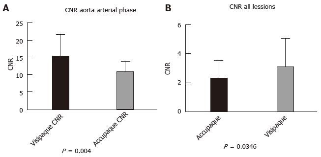
CNR of aorta to liver parenchyma on arterial phase acquisition was significantly higher for the iodixanol 270 group (15.33 ± 6.48) compared with the iohexol 350 group (10.83 ± 3.009); P = 0.0040 (Figure 1A). The comparisons between CNR for all vessels on arterial and portal vein is represented in detail in Table 1.
| Iohexol 350 group | Iodixanol 270 group | P value | ||
| Arterial Phase Acquisition | CNR Aorta/liver | 10.83 ± 3.009 | 15.33 ± 6.48 | 0.0040 |
| CNR PV/liver | 1.77 ± 1.55 | 3.40 ± 1.88 | 0.0005 | |
| CNR RPV/liver | 1.53 ± 1.87 | 2.47 ± 1.82 | 0.0129 | |
| CNR LPV/liver | 1.18 ± 1.51 | 2.36 ± 1.81 | 0.0109 | |
| Portal Phase Acquisition | CNR Aorta/liver | 3.30 ± 1.64 | 3.44 ± 1.40 | 0.7369 |
| CNR PV/liver | 4.85 ± 1.52 | 7.38 ± 2.89 | 0.0001 | |
| CNR RPV/liver | 4.49 ± 1.61 | 6.92 ± 2.58 | < 0.0001 | |
| CNR LPV/liver | 4.43 ± 2.32 | 6.82 ± 2.27 | 0.0002 | |
In total, 56 focal liver lesions (mean diameter 25.4 ± 18.9 mm) were identified in the Iohexol group and 53 (mean diameter 21.4 ± 14.5 mm) in the Iodixanol group in all three enhanced phase series. Overall CNR was significantly higher for the Iodixanol group (3.07 ± 1.96) compared to the Iohexol group (2.30 ± 1.23; P = 0.0346, Figure 1B). The analysis and comparison of CNR of focal lesions to liver parenchyma is shown on Table 2.
| Iohexol 350 group | Iodixanol 270 group | P values | |||||
| No. of lesions | Mean diameter(mm) | CNR | No. of lesions | Mean diameter (mm) | CNR | ||
| Hypervascular lesions/arterial phase | 12 | 14.8 ± 8.23 | 2.522 ± 1.699 | 17 | 18.1 ± 8.15 | 2.266 ± 1.243 | 0.8532 |
| Hypodense lesions/portal phase | 23 | 28.2 ± 19.5 | 2.483 ± 1.187 | 28 | 24.1 ± 17.8 | 3.744 ± 2.255 | 0.0194 |
| Hypodense lesions/equilibrium phase | 21 | 28.4 ± 20.9 | 1.972 ± 0.9155 | 8 | 19.3 ± 11.0 | 2.395 ± 1.202 | 0.3162 |
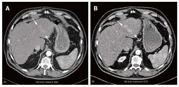
In one of the patients that were scanned with both protocols we identified one hypervascular lesion measuring 5.3 cm of greatest axis that was subjectively more evident with iodixanol 270 protocol, with a CNR of 0.36 for iohexol 350 vs 2.32 for the iodixanol 270 protocol (Figure 2).
Total radiation dose for iohexol 350 group was 22.0 ± 8.31 mSv and for iodixanol 270 group 12.9 ± 4.26 mSv. Radiation dose for unenhanced, arterial, portal and equilibrium phase acquisitions were 4.67 ± 1.97, 4.42 ± 1.88, 8.46 ± 3.07 and 2.36 ± 0.757, for the iohexol 350 group and 2.36 ± 0.757, 2.44 ± 0.923, 5.72 ± 2.27, 2.17 ± 0.756 for iodixanol 270 group, respectively.
Low kilovoltage CT protocols, have been proposed for various indications in the literature. In the domain of detection of hepatocellular carcinoma, Marin et al[14] showed that low tube voltage improves conspicuity of malignant hypervascular lesions. Lee et al[15], in another study, compared low kVp four phase CT of the liver to MRI and found that results were comparable for non-obese patients. Yanaga et al[16] tested a low Iodine load (444 mgI/kg body weight) 80 kVp CT protocol for slim patients of less than 70 kg and found it to be superior to conventional 120 kVp CT protocol with 600 mgI/kg.
The approach of choosing the DECT acquisitions allows appreciating low kilovoltage acquisition series, without any potential compromise in the overall examination quality as this 80 kVp is part of a “conventional” DECT protocol and 120 kVp weighted average series was always available for diagnostic purposes. This approach has been widely used in the literature. Altenbernd et al[17] showed that low-kVp images of DECT datasets are more sensitive in detecting hypervascular liver lesions, than the weighted average 120 kVp image series, but with a decrease in subjective image quality. With the advent of iterative reconstructions, Marin et al[18,19] showed the advantage of using iterative reconstruction when low kV is used.
In the same direction, Nakaura et al[20], compared a low iodine load (360 mgI/kg) low kilovoltage (80 kVp) CT protocol with iterative reconstructions to a standard 120 kVp protocol and found no statistically significant difference for CNR between vessels and hepatic parenchyma in arterial and portal venous phase between the two protocols.
To the best of our knowledge, the present study is the first to test feasibility of low kilovoltage CT with an isoosmolar low iodine concentration contrast agent with iterative reconstructions for liver imaging.
Our results show that for both vessels and focal hepatic lesions the overall CNR is significantly higher for the low kVp, iodixanol 270 group.
In the arterial phase, CNR was significantly higher for the iodixanol 270 group (15.33 ± 6.48 vs 10.83 ± 3.09; P = 0.0040). For hypervascular focal lesions, CNR was the same for both groups (2.522 ± 1.699 vs 2.266 ± 1.243, iohexol 350 vs iodixanol 270, P = 0.8532). In order to verify that the timing of the arterial acquisition was similar for both groups the ratio of aortic to portal venous attenuation was calculated for the arterial phase acquisitions in both groups and was found to be of no statistical difference (P = 0.8762).
For portal and delayed phase acquisitions results were more homogeneous as all CNRs for all vessels, including the branches of portal veins but also for hypodense focal lesions were significantly higher for the iodixanol 270 group.
As the measurements of CNR are equal or better for the iodixanol 270 group, even including patients weighting up to 123 kg (mean weight 82.8 ± 19.9 kg), this protocol could potentially be applied to all patients and not only to slim patients as reported in the literature[16].
A limiting factor of this study is it’s retrospective nature, with a relatively limited patients number. Consequently patients were not randomized. Despite this fact, mean patients’ weight that would be a major factor to influence CT image quality between the two groups, was very similar in both groups (81.8 ± 17.7 kg for iohexol 350 group and 82.8 ± 19.9 kg for the iodixanol 270 group; P = 0.642). Another limiting factor is that the two groups consisted mainly of different patients. Thus direct comparison with paired datasets was not feasible.
Two of the patients were scanned with both protocols. In one of them we identified one hypervascular lesion measuring 5.3 cm of greatest axis that was subjectively more evident with iodixanol 270 protocol, with a CNR of 0.36 for iohexol 350 vs 2.32 for the iodixanol 270 protocol (Figure 2).
Eighty kVp 4 phase liver CT with an isoosmolar -low iodine concentration contrast agent and iterative reconstructions is feasible and yields equal or even better results compared to a conventional 120 kVp CT protocol with a low osmolar-higher iodine concentration contrast agent. This approach would be particularly useful for patients with renal or cardiac dysfunction, but would also be of benefit for the general population, as it allows considerable decrease in radiation dose.
Dynamic contrast enhanced liver computed tomography (CT) is a highly accepted tool in the detection of hypervascular and hypovascular focal liver lesions. The use of iterative reconstructions allows lowering CT image noise and theoretically makes liver imaging in low kilovoltage settings possible. Low kilovoltage would theoretically accentuate visibility of iodine contrast medium and thus allow the use of lower concentration contrast agents. The aim of the study was to verify if the proposed low kVp, with iterative reconstructions using low iodine concentration contrast agent can be used as an alternative to conventional CT protocol. The advantage of this proposed protocol would be a considerable gain in radiation dose for the general population and also a lower iodine load for special patient populations as those with impaired renal or cardiac function.
Iso-osmolar contrast media with low iodine concentration as iodixanol 270 mg/mL can have considerable benefits, especially for patients with impaired renal or cardiac function. Low-kVp CT imaging allows a better visibility of iodine contrast, but is associated with noisier images. Iterative reconstructions have been under intensive investigation recently and have been proven to significantly reduce image noise. They have also been proven to offer the same image quality in acquisitions with lower radiation dose. Dual-energy CT (DECT) is another technical development in the domain of CT. It’s use becomes available and allows studying both low and high kilovoltage acquisitions, without any compromise in diagnostic performance.
Low kilovoltage series issuing from dual-energy CT, with or without iterative reconstructions have previously been studied in the literature. However, to the best of knowledge, the present study is the first to prove the feasibility and efficacy of low kilovoltage CT with an isoosmolar low iodine concentration contrast agent with iterative reconstructions for liver imaging.
With advancing technology, iterative reconstructions become more and more robust and accessible. Low kVp CT is also gaining place as there are vendors proposing even 70 kVp CT imaging. In this context, this study can serve as a pilot study for future projects aiming to define the most performing CT-protocol for detection of focal liver lesions.
CNR: Contrast-to-noise ratio is the objective measurement that has been chosen for documentation of CT image contrast. It is calculated with the mathematical equation CNR = (Af - Al)/N, where Af stands for attenuation value of the lesion or the vessel, Al for attenuation of liver parenchyma and N for the image noise; DECT: Dual energy CT is a CT technique that consists of synchronous imaging at low and high kilovoltages. This technique gives information on tissue characteristics based on spectral analysis. Both image series of low and high kVp can be analysed separately and also a virtual image series of 120 kVp is created based on the two acquisitions and is used for reading of the CT examinations I the clinical setting; Iterative reconstructions: Technique of reconstruction of CT raw data, allowing diminishing image noise, thus resulting to a better contrast-to-noise ratio compared with the conventional filtered back projection that is the technique that has been widely used in the past years; Iso-osmolar iodinated contrast media: Contrast media being of the same osmolality as blood (290 mOsm/kg H2O).
This manuscript reports on multi-detector CT of the liver with iodixanol 270, 80 kVp. The results and analysis of polymorphism studies are fairly well reported and appear significant.
Manuscript Source: Invited manuscript
Specialty Type: Radiology, Nuclear Medicine and Medical Imaging
Country of Origin: Switzerland
Peer-Review Report Classification
Grade A (Excellent): 0
Grade B (Very good): B, B, B
Grade C (Good): 0
Grade D (Fair): 0
Grade E (Poor): 0
P- Reviewer: Liu JY, Sureka B S- Editor: Ji FF L- Editor: A E- Editor: Wu HL
| 1. | Nyman U, Björk J, Aspelin P, Marenzi G. Contrast medium dose-to-GFR ratio: a measure of systemic exposure to predict contrast-induced nephropathy after percutaneous coronary intervention. Acta Radiol. 2008;49:658-667. [RCA] [PubMed] [DOI] [Full Text] [Cited by in Crossref: 98] [Cited by in RCA: 101] [Article Influence: 5.6] [Reference Citation Analysis (0)] |
| 2. | Nyman U, Almén T, Aspelin P, Hellström M, Kristiansson M, Sterner G. Contrast-medium-Induced nephropathy correlated to the ratio between dose in gram iodine and estimated GFR in ml/min. Acta Radiol. 2005;46:830-842. [RCA] [PubMed] [DOI] [Full Text] [Cited by in Crossref: 86] [Cited by in RCA: 88] [Article Influence: 4.2] [Reference Citation Analysis (0)] |
| 3. | Lameire NH. Contrast-induced nephropathy--prevention and risk reduction. Nephrol Dial Transplant. 2006;21:i11-i23. [RCA] [PubMed] [DOI] [Full Text] [Cited by in Crossref: 61] [Cited by in RCA: 56] [Article Influence: 2.8] [Reference Citation Analysis (0)] |
| 4. | Grynne BH, Nossen JO, Bolstad B, Borch KW. Main results of the first comparative clinical studies on Visipaque. Acta Radiol Suppl. 1995;399:265-270. [PubMed] |
| 5. | Yamashita Y, Komohara Y, Takahashi M, Uchida M, Hayabuchi N, Shimizu T, Narabayashi I. Abdominal helical CT: evaluation of optimal doses of intravenous contrast material--a prospective randomized study. Radiology. 2000;216:718-723. [RCA] [PubMed] [DOI] [Full Text] [Cited by in Crossref: 189] [Cited by in RCA: 203] [Article Influence: 7.8] [Reference Citation Analysis (0)] |
| 6. | Zamboni GA, Guariglia S, Bonfante A, Martino C, Cavedon C, Mucelli RP. Low voltage CTPA for patients with suspected pulmonary embolism. Eur J Radiol. 2012;81:e580-e584. [RCA] [PubMed] [DOI] [Full Text] [Cited by in Crossref: 19] [Cited by in RCA: 20] [Article Influence: 1.3] [Reference Citation Analysis (0)] |
| 7. | Faggioni L, Neri E, Sbragia P, Pascale R, D’Errico L, Caramella D, Bartolozzi C. 80-kV pulmonary CT angiography with 40 mL of iodinated contrast material in lean patients: comparison of vascular enhancement with iodixanol (320 mg I/mL)and iomeprol (400 mg I/mL). AJR Am J Roentgenol. 2012;199:1220-1225. [RCA] [PubMed] [DOI] [Full Text] [Cited by in Crossref: 64] [Cited by in RCA: 64] [Article Influence: 4.9] [Reference Citation Analysis (0)] |
| 8. | Nakayama Y, Awai K, Funama Y, Liu D, Nakaura T, Tamura Y, Yamashita Y. Lower tube voltage reduces contrast material and radiation doses on 16-MDCT aortography. AJR Am J Roentgenol. 2006;187:W490-W497. [RCA] [PubMed] [DOI] [Full Text] [Cited by in Crossref: 132] [Cited by in RCA: 137] [Article Influence: 6.9] [Reference Citation Analysis (0)] |
| 9. | Winklehner A, Karlo C, Puippe G, Schmidt B, Flohr T, Goetti R, Pfammatter T, Frauenfelder T, Alkadhi H. Raw data-based iterative reconstruction in body CTA: evaluation of radiation dose saving potential. Eur Radiol. 2011;21:2521-2526. [RCA] [PubMed] [DOI] [Full Text] [Cited by in Crossref: 203] [Cited by in RCA: 195] [Article Influence: 13.0] [Reference Citation Analysis (0)] |
| 10. | Yoo RE, Park EA, Lee W, Shim H, Kim YK, Chung JW, Park JH. Image quality of adaptive iterative dose reduction 3D of coronary CT angiography of 640-slice CT: comparison with filtered back-projection. Int J Cardiovasc Imaging. 2013;29:669-676. [RCA] [PubMed] [DOI] [Full Text] [Cited by in Crossref: 54] [Cited by in RCA: 55] [Article Influence: 3.9] [Reference Citation Analysis (0)] |
| 11. | Volders D, Bols A, Haspeslagh M, Coenegrachts K. Model-based iterative reconstruction and adaptive statistical iterative reconstruction techniques in abdominal CT: comparison of image quality in the detection of colorectal liver metastases. Radiology. 2013;269:469-474. [RCA] [PubMed] [DOI] [Full Text] [Cited by in Crossref: 81] [Cited by in RCA: 84] [Article Influence: 6.5] [Reference Citation Analysis (0)] |
| 12. | Singh S, Kalra MK, Do S, Thibault JB, Pien H, O’Connor OJ, Blake MA. Comparison of hybrid and pure iterative reconstruction techniques with conventional filtered back projection: dose reduction potential in the abdomen. J Comput Assist Tomogr. 2012;36:347-353. [RCA] [PubMed] [DOI] [Full Text] [Cited by in Crossref: 120] [Cited by in RCA: 114] [Article Influence: 8.1] [Reference Citation Analysis (0)] |
| 13. | Graser A, Johnson TR, Chandarana H, Macari M. Dual energy CT: preliminary observations and potential clinical applications in the abdomen. Eur Radiol. 2009;19:13-23. [RCA] [PubMed] [DOI] [Full Text] [Cited by in Crossref: 378] [Cited by in RCA: 359] [Article Influence: 19.9] [Reference Citation Analysis (0)] |
| 14. | Marin D, Nelson RC, Samei E, Paulson EK, Ho LM, Boll DT, DeLong DM, Yoshizumi TT, Schindera ST. Hypervascular liver tumors: low tube voltage, high tube current multidetector CT during late hepatic arterial phase for detection--initial clinical experience. Radiology. 2009;251:771-779. [RCA] [PubMed] [DOI] [Full Text] [Cited by in Crossref: 197] [Cited by in RCA: 183] [Article Influence: 10.8] [Reference Citation Analysis (0)] |
| 15. | Lee CH, Kim KA, Lee J, Park YS, Choi JW, Park CM. Using low tube voltage (80kVp) quadruple phase liver CT for the detection of hepatocellular carcinoma: two-year experience and comparison with Gd-EOB-DTPA enhanced liver MRI. Eur J Radiol. 2012;81:e605-e611. [RCA] [PubMed] [DOI] [Full Text] [Cited by in Crossref: 19] [Cited by in RCA: 20] [Article Influence: 1.4] [Reference Citation Analysis (0)] |
| 16. | Yanaga Y, Awai K, Nakaura T, Utsunomiya D, Funama Y, Date S, Yamashita Y. Hepatocellular carcinoma in patients weighing 70 kg or less: initial trial of compact-bolus dynamic CT with low-dose contrast material at 80 kVp. AJR Am J Roentgenol. 2011;196:1324-1331. [RCA] [PubMed] [DOI] [Full Text] [Cited by in Crossref: 36] [Cited by in RCA: 36] [Article Influence: 2.4] [Reference Citation Analysis (0)] |
| 17. | Altenbernd J, Heusner TA, Ringelstein A, Ladd SC, Forsting M, Antoch G. Dual-energy-CT of hypervascular liver lesions in patients with HCC: investigation of image quality and sensitivity. Eur Radiol. 2011;21:738-743. [RCA] [PubMed] [DOI] [Full Text] [Cited by in Crossref: 89] [Cited by in RCA: 86] [Article Influence: 5.4] [Reference Citation Analysis (0)] |
| 18. | Marin D, Nelson RC, Schindera ST, Richard S, Youngblood RS, Yoshizumi TT, Samei E. Low-tube-voltage, high-tube-current multidetector abdominal CT: improved image quality and decreased radiation dose with adaptive statistical iterative reconstruction algorithm--initial clinical experience. Radiology. 2010;254:145-153. [RCA] [PubMed] [DOI] [Full Text] [Cited by in Crossref: 419] [Cited by in RCA: 409] [Article Influence: 25.6] [Reference Citation Analysis (0)] |
| 19. | Marin D, Choudhury KR, Gupta RT, Ho LM, Allen BC, Schindera ST, Colsher JG, Samei E, Nelson RC. Clinical impact of an adaptive statistical iterative reconstruction algorithm for detection of hypervascular liver tumours using a low tube voltage, high tube current MDCT technique. Eur Radiol. 2013;23:3325-3335. [RCA] [PubMed] [DOI] [Full Text] [Cited by in Crossref: 29] [Cited by in RCA: 26] [Article Influence: 2.0] [Reference Citation Analysis (0)] |
| 20. | Nakaura T, Nakamura S, Maruyama N, Funama Y, Awai K, Harada K, Uemura S, Yamashita Y. Low contrast agent and radiation dose protocol for hepatic dynamic CT of thin adults at 256-detector row CT: effect of low tube voltage and hybrid iterative reconstruction algorithm on image quality. Radiology. 2012;264:445-454. [RCA] [PubMed] [DOI] [Full Text] [Cited by in Crossref: 109] [Cited by in RCA: 120] [Article Influence: 8.6] [Reference Citation Analysis (0)] |


