Published online Dec 28, 2016. doi: 10.4329/wjr.v8.i12.933
Peer-review started: July 31, 2016
First decision: September 2, 2016
Revised: September 19, 2016
Accepted: October 17, 2016
Article in press: October 18, 2016
Published online: December 28, 2016
Processing time: 142 Days and 19.4 Hours
This letter is a commentary on the article titled “Evaluation of variations in sinonasal region with computed tomography”, published in the January 2016 issue of World Journal of Radiology. The authors definition of the secondary middle turbinate is incorrect. The authors stated that the secondary middle turbinate is an accessory turbinate that is seen between the superior and middle turbinates. It should originate from the middle meatus posterosuperior to the ethmoid infundibulum.
Core tip: This letter is a commentary on the article titled “Evaluation of variations in sinonasal region with computed tomography,” published in the January 2016 issue of World Journal of Radiology. The authors evaluated the paranasal sinus tomography of 400 patients to determine the frequency of 39 anatomic variations. Their study required a great deal of time and effort. Unfortunately, their definition of the secondary middle turbinate and the figure that showed its structure are incorrect. It should originate, however, from the middle meatus posterosuperior to the ethmoid infundibulum, not from between the middle and superior turbinates.
- Citation: Çağıcı CA. Commentary on: “Evaluation of variations in sinonasal region with computed tomography”. World J Radiol 2016; 8(12): 933-934
- URL: https://www.wjgnet.com/1949-8470/full/v8/i12/933.htm
- DOI: https://dx.doi.org/10.4329/wjr.v8.i12.933
I read the article by Dasar et al[1] with great interest. The authors evaluated the paranasal sinus tomography of 400 patients to determine the frequency of each of 39 possible anatomic variations. Their study required a great deal of time and effort. Unfortunately, their definition of the secondary middle turbinate and the figure that showed its structure are incorrect.
The authors stated that the secondary middle turbinate is an accessory turbinate that is seen between the superior and middle turbinates[1]. It should originate, however, from the middle meatus posterosuperior to the ethmoid infundibulum, not from between the middle and superior turbinates[2]. The secondary middle turbinate is not part of the middle turbinate. The appearance of a secondary middle turbinate is probably due to the partial absence of the anterior wall of the ethmoidbulla[2].
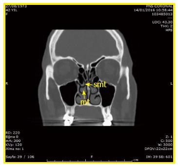
Their figure 3D, which was used to illustrate the secondary middle turbinate, is also not appropriate. A sagittal cleft on the middle turbinate is seen in this figure, which is not an appropriate example for the secondary middle turbinate. The secondary middle turbinate is actually a bony prominence that extends from the lateral nasal wall to the middle meatus, as shown in Figure 1[2].
Manuscript source: Unsolicited manuscript
Specialty type: Radiology, nuclear medicine and medical imaging
Country of origin: Turkey
Peer-review report classification
Grade A (Excellent): A, A
Grade B (Very good): B, B
Grade C (Good): C, C
Grade D (Fair): 0
Grade E (Poor): 0
P- Reviewer: Chandra R, Lobo D, Rapidis AD, Shen J, Sali L, van Beek EJR S- Editor: Kong JX L- Editor: A E- Editor: Wu HL
| 1. | Dasar U, Gokce E. Evaluation of variations in sinonasal region with computed tomography. World J Radiol. 2016;8:98-108. [RCA] [PubMed] [DOI] [Full Text] [Full Text (PDF)] [Cited by in CrossRef: 24] [Cited by in RCA: 30] [Article Influence: 3.0] [Reference Citation Analysis (0)] |
| 2. | Khanobthamchai K, Shankar L, Hawke M, Bingham B. The secondary middle turbinate. J Otolaryngol. 1991;20:412-413. [PubMed] |


