Published online Aug 28, 2014. doi: 10.4329/wjr.v6.i8.625
Revised: April 3, 2014
Accepted: July 12, 2014
Published online: August 28, 2014
Processing time: 197 Days and 17.8 Hours
Dual-energy computed-tomography (DECT) has been suggested as the method of choice for imaging urinary calculi due to the modality’s high sensitivity for detecting stones and its capability of accurately differentiating between uric-acid (UA) and non-UA (predominantly calcium) stones. The clinical significance of the latter feature relates to the differences in management of UA vs non-UA calculi. Like calculi, ureteral stents are assigned color by the dual-energy post-processing algorithm, which may lead to improved or worsened stone visualization based on the resulting stent/stone contrast. Herein we depict the case of a nephrolithiasis patient with bilateral stents, each with different color, clearly displaying the effect of stent color on stone visualization. Further, three-dimensional reconstruction of the DECT images illustrates advantages of this enhancement compared to conventional two-dimensional computed tomography. The resulting stent/stone contrast produces an unanticipated potential advantage of DECT in patients with urolithiasis and stents and may promote improved management decision-making.
Core tip: Dual-energy computed-tomography is a recently introduced technique for imaging kidney stones. Ureteral stents are also characterized by the dual-energy (DE) algorithm and, like calculi, are assigned color. The resulting stent/stone color contrast may lead to either improved or worsened stone visualization in patients with urolithiasis and stents. This case report illustrates different DE characterization of stents based on their type, and the resulting effects on stent/stone contrast. The awareness of the DE characterization of different stent types allows for stent selection that improves stone visualization.
- Citation: Ibrahim ESH, Haley WE, Jepperson MA, Wehle MJ, Cernigliaro JG. Characterization of ureteral stents by dual-energy computed tomography: Clinical implications. World J Radiol 2014; 6(8): 625-628
- URL: https://www.wjgnet.com/1949-8470/full/v6/i8/625.htm
- DOI: https://dx.doi.org/10.4329/wjr.v6.i8.625
Dual-energy computed-tomography (DECT) is a recently introduced technique for imaging urinary calculi. This technology utilizes the material change in attenuation between two peak X-ray energies to create color-coded material-specific images; in the case of urinary calculi, it is capable of distinguishing uric acid (UA) from non-UA stones[1-3]. Prior to the introduction of this technology, urinary stone composition could not be reliably determined except by attaining stone material for analysis[4,5]. The clinical significance of pre-intervention determination of stone composition lies in the fact that medical dissolution therapy may be helpful in the case of UA calculi, and may obviate the need for surgical procedures that are often required for treatment of non-UA calculi[6,7].
In cases of ureteral obstruction or procedures involving the ureter, stent placement is commonly required. Ureteral stents are also characterized by the dual-energy (DE) algorithm; those with similar attenuation change as UA stones are assigned one color (e.g., red) while those with change in attenuation similar to non-UA stones are assigned another color (e.g., blue). We have previously reported a list of the DE characteristics of commonly-used ureteral stents[8]. The resulting potential for stent/stone color contrast creates an unanticipated advantage of DECT in patients with urolithiasis and stents. The stent/stone contrast may either lead to enhanced (the stent and stone have different colors) or camouflaged (both stent and stone have the same color) stone visualization. In this report, we present, to our knowledge for the first time, an interesting case of a patient with bilateral stents characterized by DECT with different colors, for whom DECT helped identify the calculi and stone composition.
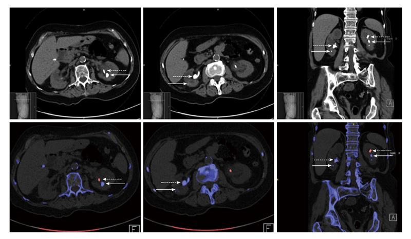
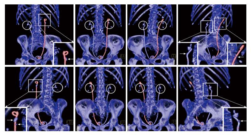
The protocol for conducting this study was approved by the Mayo Clinic institutional review board. A 78-year-old female presented with bilateral ureteral stents following ureteroscopic removal of obstructing left-side ureteral calculus, and prior stenting on right-side for obstructing renal pelvis calculus (left-side stent: Boston-Scientific; right-side stent: Cook-Amplatz). The images were acquired on a Definition-Flash Siemens computed tomography (CT) scanner in dual-energy mode. The DECT peak tube potentials (kVp) were set to 80 kV and 140 kV, and the scan was conducted using a dedicated renal stone imaging protocol. Continuous non-contrast images were obtained from the level of the diaphragms to the pubic symphysis. The data was reconstructed on a workstation provided by the manufacturer using the Syngo software (VE 36A). Axial and coronal reconstructions were made using a mixed (low and high kVp) dataset with 5 mm slice-thickness/2.5 mm slice-interval (axial) and 3 mm slice-thickness/2.5 mm slice-interval (coronal). Material-specific image reconstructions in the axial- and coronal-planes were made at 1 mm slice-thickness/0.8 mm slice-interval and D30f convolution kernel. The DE post-processing algorithm assigns color based on the ratio of X-ray attenuations from the two tube potentials using a 3-material (UA, calcium, urine) decomposition algorithm. The capability of DECT to characterize (and color code) the stones and stents is appreciated when conventional CT images are compared to the DECT characterized images, as shown in Figure 1. Note also the limitation of the two-dimensional images in Figure 1 as they show only parts of the stents and stones that lie inside the imaging plane; this is better resolved by three-dimensional image reconstruction as shown in Figure 2.
The scan revealed a 1 cm calculus in the posterior interpolar region of the left kidney and a 9 mm calculus in the left anterior lower pole calyx, as well as several nonobstructing calculi bilaterally. The dual-energy characterization showed all calculi to be of non-UA composition (blue color), while the left and right stents appeared red and blue, respectively. While the images showed excellent stent/calculus color contrast in the left kidney (stent appears red and calculi appear blue), there was virtually no observed color contrasting in the right kidney (both stent and calculi appear blue). The effect of the stent/stone contrast on calculus visualization is appreciated when the images are viewed from different angles as illustrated in the three-dimensional reconstructed images in Figure 2, where calculus in the right kidney is totally camouflaged by the stent when viewed from certain angles.
DECT scanners acquire data simultaneously from two X-ray tubes (operating at different potentials) and detector arrays that are mounted near perpendicular angles[1,2]. DECT is advantageous compared to conventional CT for nephrolithiasis imaging due to the modality’s additional capability of characterizing the stone composition a priori and without increasing radiation dose, which allows for optimizing the treatment decision-making (DECT is nearly 100% accurate for distinguishing UA from non-UA stones > 3 mm)[4-6]. Nevertheless, the situation is more complicated in patients requiring stents, as they appear colored in the resulting images based on their individual DE characterization[8]. Herein, we depict an interesting and illustrative case with bilateral stents that were assigned different colors in the resulting DECT images (Boston Scientific stent left ureter and Cook-Amplatz stent right ureter). Stent coloring may be an advantage when the stent and calculus are assigned different colors, as the stone is better recognized with better appreciation of its size and shape. On the other hand, the situation may be worsened when both stent and stone are assigned the same color, as the stent may conceal or camouflage the stone and/or its boundary. In the latter case, premature stent removal in the presence of potential obstructing stone fragments could follow. Accordingly, awareness of the DE characterization of different stent types allows for appropriate stent selection in patients with known stone composition. The potential for stent/stone color contrast connotes a possible advantage of DECT in patients with urolithiasis requiring a stent and may thereby promote improved management decision-making.
A 78-year-old kidney stone female patient with bilateral ureteral stents.
Nephrolithiasis with dysuria, hematuria, and bilateral flank pain.
Acute kidney injury with bilateral obstructing stones requiring bilateral stents.
Lab results showed serum creatine of 2.8 mg/dL; urine culture: > 100000 cfu/mL Escherichia coli; urinalysis: > 182 wbc/hpf, > 182 rbc/hpf, pH 5.5, 2+ protein.
Bilateral moderate hydronephrosis and hydroureter and multiple large renal calculi bilaterally: large right renal pelvic stone 2 cm × 1.6 cm × 1.2 cm with dilated calyces distally; 7 mm ureteral calculus mid/distal left ureter.
Stone analysis by infrared spectroscopy revealed stone composition of 90%/10% of calcium oxalate monohydrate/calcium phosphate (apatite).
The patient underwent ureteroscopic removal of obstructing left-side ureteral calculus with stent placement as well as stent placement on the right side for obstructing renal pelvis calculus.
Dual-energy computed-tomography (DECT) is capable of distinguishing uric acid (UA) from non-UA stones; moreover, a recent report provided dual-energy characteristics of commonly used ureteral stents.
DECT is a recently introduced technique for imaging urinary calculi, whereby the material change in attenuation between two peak X-ray energies is used to create color-coded material-specific images.
This case report shows the potential advantage of DECT in patients with urolithiasis requiring a stent, where selection of the stent type based on its dual-energy characteristics improves the resulting stent/stone color contrast for better management decision-making.
This report shows the capability of DECT to distinguish kidney stones by color-coding the stones and ureteral stents as compared to conventional computed tomography. This technology is novel and useful in determining the stone type, a distinction that is important for determining the optimal treatment strategy.
| 1. | Matlaga BR, Kawamoto S, Fishman E. Dual source computed tomography: a novel technique to determine stone composition. Urology. 2008;72:1164-1168. [RCA] [PubMed] [DOI] [Full Text] [Cited by in Crossref: 90] [Cited by in RCA: 78] [Article Influence: 4.3] [Reference Citation Analysis (0)] |
| 2. | Hartman R, Kawashima A, Takahashi N, Silva A, Vrtiska T, Leng S, Fletcher J, McCollough C. Applications of dual-energy CT in urologic imaging: an update. Radiol Clin North Am. 2012;50:191-205, v. [RCA] [PubMed] [DOI] [Full Text] [Cited by in Crossref: 46] [Cited by in RCA: 40] [Article Influence: 2.9] [Reference Citation Analysis (0)] |
| 3. | Boll DT, Patil NA, Paulson EK, Merkle EM, Simmons WN, Pierre SA, Preminger GM. Renal stone assessment with dual-energy multidetector CT and advanced postprocessing techniques: improved characterization of renal stone composition--pilot study. Radiology. 2009;250:813-820. [RCA] [PubMed] [DOI] [Full Text] [Cited by in Crossref: 185] [Cited by in RCA: 160] [Article Influence: 9.4] [Reference Citation Analysis (0)] |
| 4. | Kulkarni NM, Eisner BH, Pinho DF, Joshi MC, Kambadakone AR, Sahani DV. Determination of renal stone composition in phantom and patients using single-source dual-energy computed tomography. J Comput Assist Tomogr. 2013;37:37-45. [RCA] [PubMed] [DOI] [Full Text] [Cited by in Crossref: 71] [Cited by in RCA: 67] [Article Influence: 5.2] [Reference Citation Analysis (0)] |
| 5. | Ascenti G, Siragusa C, Racchiusa S, Ielo I, Privitera G, Midili F, Mazziotti S. Stone-targeted dual-energy CT: a new diagnostic approach to urinary calculosis. AJR Am J Roentgenol. 2010;195:953-958. [RCA] [PubMed] [DOI] [Full Text] [Cited by in Crossref: 76] [Cited by in RCA: 77] [Article Influence: 4.8] [Reference Citation Analysis (0)] |
| 6. | Stolzmann P, Kozomara M, Chuck N, Müntener M, Leschka S, Scheffel H, Alkadhi H. In vivo identification of uric acid stones with dual-energy CT: diagnostic performance evaluation in patients. Abdom Imaging. 2010;35:629-635. [RCA] [PubMed] [DOI] [Full Text] [Cited by in Crossref: 85] [Cited by in RCA: 81] [Article Influence: 4.8] [Reference Citation Analysis (0)] |
| 7. | Primak AN, Fletcher JG, Vrtiska TJ, Dzyubak OP, Lieske JC, Jackson ME, Williams JC, McCollough CH. Noninvasive differentiation of uric acid versus non-uric acid kidney stones using dual-energy CT. Acad Radiol. 2007;14:1441-1447. [PubMed] |
| 8. | Jepperson MA, Thiel DD, Cernigliaro JG, Broderick GA, Parker AS, Haley WE. Determination of ureter stent appearance on dual-energy computed tomography scan. Urology. 2012;80:986-989. [RCA] [PubMed] [DOI] [Full Text] [Cited by in Crossref: 9] [Cited by in RCA: 9] [Article Influence: 0.6] [Reference Citation Analysis (0)] |
P- Reviewer: Sakhaee K S- Editor: Wen LL L- Editor: A E- Editor: Liu SQ


