INTRODUCTION
Differentiated thyroid carcinoma (DTC) comprises the majority of thyroid cancers and usually is indolent with a good prognosis and survival rate. However, in a fraction of cases the disease could behave aggressively with the presence of distant metastases at diagnosis. We herein report a rare case of follicular thyroid carcinoma (FTC) with adrenal metastasis. FTC is a histological subtype of differentiated thyroid carcinoma which is more aggressive than papillary thyroid carcinoma (PTC) due to its propensity of hematogenous spread. The most common mode of spread of FTC is by vascular invasion with metastases commonly seen in the skeletal system and lungs. However, there have been occasional reports of metastases to other soft tissue such as the brain, kidneys, liver and adrenals[1-17].
CASE REPORT
A 55-year-old female patient presented with a painless anterior neck swelling and a sternal mass. An initial evaluation by contrast enhanced computed tomography (CT) scan demonstrated a bulky right lobe of the thyroid with a hypodense lesion (2.2 cm × 2.1 cm) associated with a peripheral rim of calcification. A lytic sternal lesion along with a large extra-osseous soft tissue mass (9 cm × 7 cm) showed heterogeneous enhancement with a central necrotic area and intra-thoracic extension. Multiple soft tissue density nodules were also observed in bilateral lung parenchyma. Fine needle aspiration cytology (FNAC) from the thyroid lesion was suggestive of a follicular variant of papillary thyroid carcinoma. FNAC from the sternal lesion showed pattern of microfollicular carcinoma.
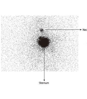
The patient underwent total thyroidectomy with the histopathology confirmatory of differentiated widely invasive follicular carcinoma of thyroid. After surgery she was referred for an I-131 scan and subsequent I-131 therapy. A post surgery 3.7 MBq I-131 scan (Figure 1) revealed radioiodine concentration in the neck region (24 hour neck uptake - 3.39%) and over the sternal region (uptake - 19.44%). She was administered 7.4 GBq of I-131 for therapeutic purposes. Post-radioiodine therapy (PT scan) (Figure 2) revealed radioiodine uptake in the thyroid bed, sternum and a focus of intense radioiodine concentration in the left suprarenal region. The lesion seen in the left suprarenal region was not seen in the initial CT evaluation as the CT scan was limited to the neck and chest region. Spot oblique images and single photon emission computed tomography (SPECT) of the upper abdomen (Figure 3) were taken to ascertain the position of the lesion. A subsequent whole body PET-CT (non-contrast) revealed a large fluorodeoxyglucose (FDG) concentrating heterogeneous mass in the left adrenal gland region in addition to a FDG avid osteolytic sternal lesion and multiple FDG avid bilateral pulmonary nodules (Figure 4). The thyroglobulin estimated by immunoradiometric assay technique was > 250 ng/mL with thyroid stimulating hormone thyroid stimulating hormone (TSH) of 4.9 μIU/mL, even following the levothyroxine withdrawal protocol, suggesting a functioning nature of the metastases. During the writing this report, the patient is doing well on follow up, in spite of extensive metastasis, and is ambulatory. She is due for the next radio-iodine therapy in 2 mo.
Figure 1 Pre-treatment diagnostic radioiodine scan (post thyroidectomy at 5 wk) demonstrating neck uptake and iodine avid sternal lesion.
No other abnormal focus was noted elsewhere in the body.
Figure 2 Post therapy scan while discharging the patient from radioiodine therapy ward demonstrates a left abdominal focal lesion in addition to the neck and sternal focus.
Figure 3 Post-therapy scan demonstrating the left sided abdominal focus more vividly.
A: Planar spot view of abdomen; B: Single-photon emission computed tomography (SPECT) coronals (SPECT images); C: The 3-slice SPECT display (transaxials, sagittals, coronals): Arrow indicates the adrenal metastasis.
Figure 4 Positron emission tomography-computed tomography fusion images demonstrating avid fludeoxyglucose uptake in the left adrenal lesion.
A: Transaxial fludeoxyglucose-PET/CT (CT, PET, fused PET-CT); B: Fused Coronals. PET/CT: Positron emission tomography/computed tomography.
DISCUSSION
Around 10% of patients with DTC present with multiple sites of distant metastasis such as the lung, bone and lymph nodes. Among the other sites of metastases, the brain comprises 50%, liver 25% and other sites 25%[1]. To date, 11 cases of adrenal metastasis secondary to differentiated thyroid carcinoma have been reported in the literature[2]. They are usually asymptomatic and frequently detected by the post-therapy scan. Detection of rare soft tissue metastases through different diagnostic modalities has been described in multiple case reports in the literature. A variable expression of sodium iodide symporter among different metastatic sites has also been described which could be one of the reasons for which such metastases (including the adrenal metastasis) are only rarely detected, at times at autopsy[3-5].
Out of 11 documented cases of adrenal metastases, 5 arose in the setting of follicular carcinoma, 4 in PTC and 2 were from Hurthle cell variant. Most of the reported cases of adrenal metastasis were asymptomatic, as was observed in our case[6-11]. Such adrenal metastatic lesions in the setting of differentiated thyroid carcinoma are quite rare[12-17]. These metastases are generally associated with lung and skeletal metastases and their detection is difficult even if they are iodine concentrating owing to their proximity of the physiological uptake of radio-iodine in the gut or their excretion route through kidney.
Our case had the histology of widely invasive differentiated follicular carcinoma. There was an intense and focal abdominal radio-iodine uptake, as documented on planar and SPECT images, leading to suspicion and detection of adrenal metastasis in this patient. Even in the absence of availability of SPECT-CT, intense focal radio-iodine uptake or uptake associated with star effect in the upper abdomen needs to be closely investigated to differentiate it from physiological radio-iodine concentration in the gut, biliary or urinary tract which is usually more diffuse and rarely gives star artifact[18]. The location of any radio-iodine activity in planar imaging should be further characterized as anterior and posterior depending upon the intensity on anterior and posterior projections. Lateral and oblique views may also be of substantial value and be obtained to distinguish adrenal uptake, which will be located posteriorly from gastric or hepatic uptake. A SPECT imaging can subserve a valuable purpose in diagnosing the suspected area and thus should be mandatorily obtained in the presence of a suspicious focus[19]. However, one has to interpret and further evaluate any such findings against the possibility of false positive findings, as in the case of radio-iodine concentration in a renal cyst and biliary tract[20-21]. Hence, in the presence of a positive post therapy radio-iodine concentration scan with focal radio-iodine concentration in the abdomen, a high index of suspicion of metastases to an intra abdominal organ should be raised and needs to be effectively excluded by anatomical imaging (CT/magnetic resonance imaging/ultrasonography) or SPECT/CT hybrid imaging. A FDG-PET/CT performed in our case further confirmed the lesion in the supra renal region and documented a well defined 6.5 cm × 5.0 cm adrenal lesion with a SUVmax (standardized uptake value-maximum) of 9.5. In our patient with Tg > 250 ng/mL and TSH of 4.9 μΙU/mL, the functioning nature of the metastatic lesions was indicated, even after thyroid hormone withdrawal. In functioning metastases, even with ensuring adequate withdrawal of levothyroxine, the TSH level could be generally within the normal range as seen in the present case. The primary aim of radio-iodine treatment in these patients, of course, is disease stabilization, symptom palliation with prolongation of symptom free survival and improvement in quality of life. While in some reports large volume distant metastatic lesion has also been managed with surgery, particularly in the setting of skeletal metastases[22-25], our patient opted for radio-iodine treatment and refused surgery and hence was treated with the same. The other important feature that can be deciphered from this report of adrenal metastasis is that it could be unilateral and solitary, unlike those of renal metastases which are almost always bilateral and multiple at presentation, although both are usually asymptomatic[4,5].
In conclusion, patients with differentiated thyroid carcinoma may be asymptomatic even if harboring unlikely metastatic sites such as the adrenals, which are seen along with other sites of metastatic disease such as pulmonary or skeletal metastasis. A high degree of clinical suspicion coupled with careful clinicoradiological correlation by other imaging modalities could prove invaluable in the detection of such rare metastatic lesions. The importance of a post-therapy radio-iodine scan in the detection of unidentified metastatic lesions (during initial pre-treatment diagnostic study) can hardly be overemphasized.
COMMENTS
Case characteristics
A 55-year-old female patient presented with a painless anterior neck swelling and a sternal mass.
Clinical diagnosis
Both sternal and anterior neck swellings were hard in consistency. Anterior neck swelling moves with deglutition, indicating thyroid pathology.
Laboratory diagnosis
white blood cell 7.40 k/uL; haemoglobin11.5 gm/dL; TG > 250 ng/mL; metabolic panel and renal function tests were within normal limits.
Pathological diagnosis
Fine needle aspiration cytology (FNAC) from the thyroid lesion was suggestive of a follicular variant of papillary carcinoma thyroid. FNAC from the sternal lesion showed a pattern of microfollicular carcinoma.
Treatment
The patient underwent total thyroidectomy. After surgery 7.4 GBq of I-131 was administered for therapeutic purposes.
Related reports
Differentiated thyroid carcinoma metastasizing to adrenals is rare, with only 11 such documented cases.
Term explanation
A post therapy I-131 scan is a scan done after administration of a therapeutic dose of I-131 in differentiated thyroid carcinoma to see the sites where the radio-iodine has localized and to find any occult metastatic sites missed out on the initial evaluation.
Experiences and lessons
This case report not only represents the rareness of adrenal metastasis in cases of differentiated thyroid carcinoma, but also emphasizes the importance of a post therapy scan in detecting any occult metastasis not diagnosed on initial evaluation.
Peer review
Adrenal metastases from differentiated thyroid cancers (DTC) are exceptional. Very interesting case on a unilateral adrenal metastasis from a follicular DTC detected on a post-therapy 131-I scan.


