Published online May 28, 2021. doi: 10.4329/wjr.v13.i5.137
Peer-review started: February 19, 2021
First decision: March 28, 2021
Revised: March 31, 2021
Accepted: May 22, 2021
Article in press: May 22, 2021
Published online: May 28, 2021
Processing time: 97 Days and 21.3 Hours
Few systematic comparative studies of the different methods of physical elastography of the spleen are currently available.
To compare point shear wave and two-dimensional elastography of the spleen considering the anatomical location (upper, hilar, and lower pole).
As part of a prospective clinical study, healthy volunteers were examined for splenic elasticity using four different ultrasound devices between May 2015 and April 2017. The devices used for point shear wave elastography were from Siemens (S 3000) and Philips (Epiq 7), and those used for two-dimensional shear wave elastography were from GE (Logiq E9) and Toshiba (Aplio 500). In addition, two different software versions (5.0 and 6.0) were evaluated for the Toshiba ultrasound device (Aplio 500). The study consisted of three arms: A, B, and C.
In study arm A, 200 subjects were evaluated (78 males and 122 females, mean age 27.9 ± 8.1 years). In study arm B, 113 subjects were evaluated (38 men and 75 women, mean age 26.0 ± 6.3 years). In study arm C, 44 subjects were enrolled. A significant correlation of the shear wave velocities at the upper third of the spleen (r = 0.33088, P < 0.0001) was demonstrated only for the Philips Epiq 7 device compared to the Siemens Acuson S 3000. In comparisons of the other ultrasound devices (GE, Siemens, Toshiba), no comparable results could be obtained for any anatomical position of the spleen. The influencing factors age, gender, and body mass index did not show a clear correlation with the measured shear wave velocities.
The absolute values of the shear wave elastography measurements of the spleen and the two different elastography methods are not comparable between different manufacturers or models.
Core Tip: Measurement of shear wave velocity in the spleen has been increasingly used in prognostic assessment of esophageal varices and as a marker of portal hypertension. The current recommendations of medical societies for splenic elastography note methodological limitations in transient elastography. Currently, whether the different elastography methods and shear wave measurements with different ultrasonic devices provide comparable results has not been clarified. Our results show that the most reliable measurements for all devices were obtained at the lower pole of the spleen. However, absolute values of splenic shear wave elastography measurements are not transferable between manufacturers or models.
- Citation: Nowotny F, Schmidberger J, Schlingeloff P, Binzberger A, Kratzer W. Comparison of point and two-dimensional shear wave elastography of the spleen in healthy subjects. World J Radiol 2021; 13(5): 137-148
- URL: https://www.wjgnet.com/1949-8470/full/v13/i5/137.htm
- DOI: https://dx.doi.org/10.4329/wjr.v13.i5.137
Ultrasound shear wave elastography is gaining importance in diagnostics for a variety of diseases[1-4]. In recent years, several ultrasound-based elastography techniques have been developed for non-invasive quantitative assessment of tissue elasticity, primarily liver stiffness[5]. The first method in this field was transient elastography (TE) by FibroScan. A newer generation of elastography techniques that do not require mechanical pulses to generate shear waves, but instead use high-intensity ultrasound waves, is summarized as acoustic radiation force impulse (ARFI) elastography. Compared to TE, ARFI techniques are more precise and allow more valid measurements, even in patients with high body mass index (BMI) or ascites[6].
Currently, there are two different techniques that work on the basis of this principle, both of which can be generally summarized under the term shear wave elastography: point shear wave elastography (pSWE) and two-dimensional shear wave elastography (2D-SWE)[5]. Different manufacturers have increasingly integrated p-SWE and 2D-SWE techniques into their ultrasound scanners. In a meta-analysis, the pSWE and 2D-SWE techniques showed significantly better results than FibroScan with respect to the rate of unreliable measurements in healthy subjects and in patients with chronic liver disease[6]. However, some of the study populations examined in the comparative studies were small and, often, only two different ultrasound devices from different manufacturers were compared[7-9]. Recent studies with larger samples have shown good agreement between the p-SWE and 2D-SWE techniques for different manufacturers, with slightly lower shear wave velocities for 2D-SWE depending on the software version used[10-12]. Furthermore, various factors, such as fasting time, breathing, and BMI, can substantially affect the measurement of shear wave velocities and, therefore, must be taken into account when interpreting the results[5]. A recent meta-analysis of 2D-SWE reconfirmed the higher reliability of the method compared to the other ultrasound elastography methods as demonstrated in various studies[6].
In recent years, the measurement of splenic stiffness has increasingly become the focus of scientific investigations, especially for prognostic assessment of esophageal varices and as a marker of portal hypertension[13-15]. In a recent meta-analysis, Song et al[16] demonstrated a good correlation between splenic stiffness and blood pressure measured by hepatic venous pressure gradient. The current recommendations of the European Federation of Societies for Ultrasound in Medicine and Biology (EFSUMB) note obvious methodological limitations for transient splenic ultrasonography, especially in patients with high BMI, the detection of ascites and pulmonary or colonic gas overlays, and in patients with a splenic diameter < 4 cm. The successful application of TE in measuring splenic elasticity has been reported to be approximately 70%[15]. With few studies currently available on 2D-SWE, the technology has been viewed critically in the assessment of splenic stiffness[15,17]. For p-SWE, recent studies report sensitivity of up to 97% for the measurement of splenic stiffness, but spleen size, adiposity, and abdominal wall thickness seem to affect reproducibility in p-SWE[15,18-21]. In addition to the above parameters, the anatomical position for measurement in splenic elastography seems to influence the results. To date, most elastography studies on the spleen have performed measurements at undefined anatomic positions or different splenic poles[22-26].
To the best of our knowledge, no comparative studies are currently available on different elastography methods (e.g., pSWE, 2D-SWE) for sonoelastographic measurement of splenic stiffness taking into account the anatomical location of the measurement in healthy volunteers. The aim of the present study was to compare 2D-SWE to p-SWE in healthy volunteers taking into account whether the measurement is performed in the upper, middle, or lower third of the spleen.
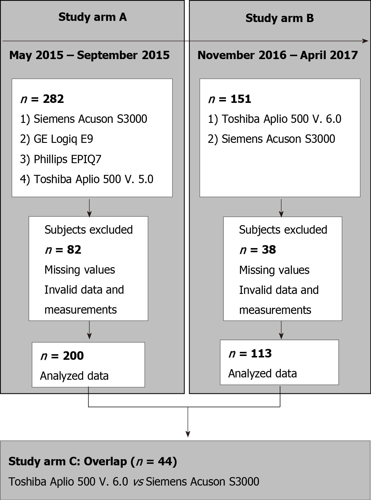
The study consisted of three arms: A, B, and C (Figure 1). Arm A tested four ultrasound devices: the Siemens Acuson S3000, Toshiba Aplio 500 (software version 5.0), Philips Epiq 7, and GE Logiq E9. We chose the Siemens S3000 ultrasound scanner as the reference device because the largest number of studies exist for this ultrasound scanner or its predecessor, the Siemens S2000[27-29]. The Siemens S3000 and Philips Epiq 7 devices use p-SWE technology, and the Aplio 500 Toshiba and GE Logiq E9 devices use 2D-SWE technology. Study arm A showed that the Toshiba Aplio 500 device (software version 5.0) generated strongly deviating results compared to the other ultrasound devices tested. Due to the divergent results between Toshiba Aplio 500 (version 5.0) and the other tested devices, especially the reference device, the Toshiba Aplio 500 was tested using software version 6.0 against the Siemens Acuson S3000 in study arm B. In study arm C, the results of study arms A and B were compared to investigate the differences between the two different software versions of the Toshiba Aplio 500. Study arm A was conducted from May 2015 to September 2015 and study arm B from November 2016 to April 2017.
Initially, 282 subjects were included in study arm A. Due to incomplete measurements and invalid data and measurements, the data sets of 200 subjects could be evaluated. In study arm B, 151 subjects were initially recruited, but because of missing or incomplete data 113 subjects could be analyzed. The characteristics of the subjects in study arms A and B are given in Table 1. Study arm C included 44 subjects. The same study protocol applied to both study arms. Only subjects who met the inclusion criteria and provided informed written consent to participate in the study were recruited. The study had a positive vote from the local ethics committee (No. 415/15) and was conducted according to the guidelines of the Declaration of Helsinki[30]. The inclusion criteria in the study were age ≥ 18 years; no history of hepatopathies (viral hepatides, hemacromatosis, autoimmune hepatitis, toxic hepatides, Wilson's disease) or other chronic diseases, such as diabetes or arterial hypertension; fasting period ≥ 3 h before ultrasound examination; BMI < 30 kg/m² and > 18 kg/m²; normal findings on previous abdominal ultrasonography, specifically normal echogenicity, texture, and size of the liver (≤ 16 cm) and normal echogenicity and size of the spleen (up to 14 cm length allowed); and alcohol consumption < 40 g/d in men and < 20 g/d in women.
| Study arm A (n = 200) | Study arm B (n = 113) | Overlap C (n = 44) | ||||
| Frequency | mean ± SD | Frequency | mean ± SD | Frequency | mean ± SD | |
| Gender | ||||||
| Male | 78 (39.00) | 38 (30.6) | 10 (22.73) | |||
| Female | 122 (61.00) | 75 (66.4) | 34 (77.27) | |||
| Age | 27.93 ± 8.13 | 25.95 ± 6.26 | 27.11 ± 5.64 | |||
| < 30 yr | 154 (77.00) | 98 (86.7) | 34 (77.27) | |||
| ≥ 30 yr | 46 (23.00) | 15 (13.3) | 10 (22.73) | |||
| BMI | 22.56 ± 2.57 | 21.64 ± 2.24 | 21.60 ± 2.67 | |||
| BMI < 25 | 166 (83.0) | 103 (91.2) | 38 (86.36) | |||
| BMI ≥25 | 34 (17.00) | 10 (8.9) | 6 (13.64) | |||
| Alcohol consumption | 8.17 ± 9.70 | 6.73 ± 8.35 | 6.22 ± 7.95 | |||
| None | 18 (9.00) | 9 (8.0) | 9 (20.45) | |||
| Less 1/mo | 28 (14.00) | 17 (15.0) | 3 (6.82) | |||
| Several times per month | 106 (53.00) | 71 (62.8) | 28 (63.64) | |||
| Several times per week | 48 (24.00) | 16 (14.2) | 4 (9.09) | |||
| Fasting time | 3.74 ± 1.84 | 4.47 ± 2.80 | 4.11 ± 1.65 | |||
| Spleen length (mm) | 105.44 ± 14.80 | 105.53 ± 14.16 | 101.66 ± 13.78 | |||
| Median (min-max) | 105.50 (61-144) | 106.00 (65-144) | 100.50 (75-137) | |||
| Spleen depth (mm) | 35.38 ± 6.19 | 35.28 ± 6.38 | 34.25 ± 5.59 | |||
| Median (min-max) | 35.50 (21-56) | 35.00 (21-56) | 34.00 (24-48) | |||
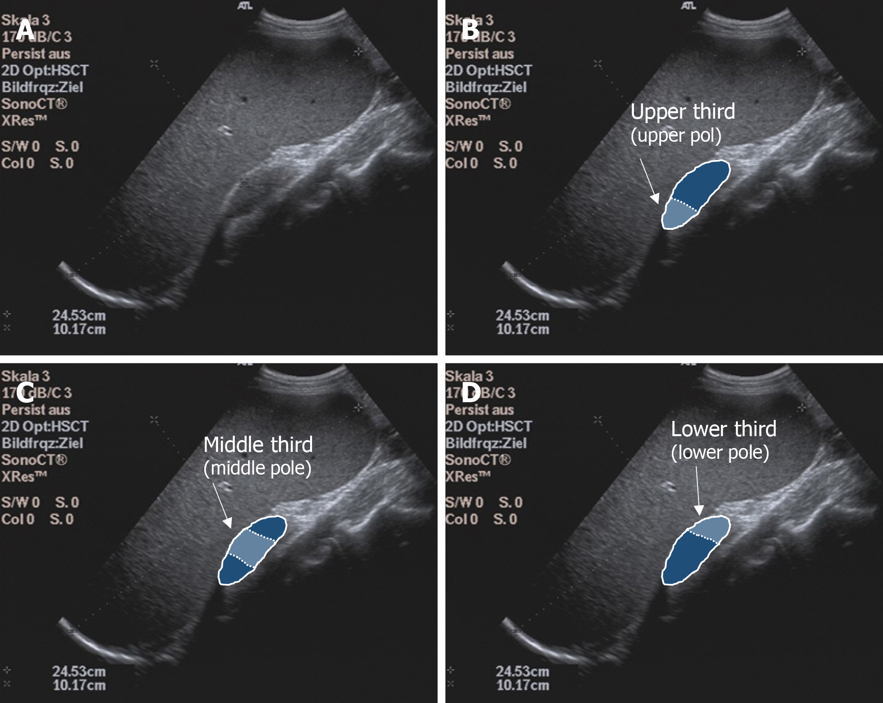
Before elastography, standardized abdominal ultrasonography of the liver and spleen in B-mode was performed in each subject to document the liver size, echogenicity, and parenchymal structure and the spleen size, shape, and parenchymal and vascular status. Subjects with pathological findings on focused abdominal ultrasonography were excluded from the study. One subject at a time was examined by one investigator using all devices. Study arm A had 6 investigators and study arm B had 2 investigators; an experienced supervisor (> 5000 examinations/year) was available in case of unclear findings. Splenic elastography was performed in all subjects in the supine position with the left arm maximally abducted and in expiration. Care was taken to place the transducer at right angles to the splenic capsule as much as possible. Shear wave velocity measurements were obtained in meters per second at each of three anatomic positions: the upper, middle, and lower thirds of the spleen (Figure 2). Five valid measurements were obtained per anatomic position using the Philips Epiq 7 and Siemens Acuson S3000 to calculate a median and mean value. A total of 15 measurements per spleen were performed using p-SWE. Elastographic studies on the Toshiba Aplio 500, GE Logiq E9, and Siemens Acuson S3000 were performed with convex transducers (6C1HD, 1.5-5.5 MHz) and on the Philips Epiq 7 with one transducer (5C1 HD, 1-5 MHz). The preset region of interest (ROI) was 10 mm × 5 mm for Siemens. The ROI for the other manufacturers was set to 10 mm × 10 mm. As the quality of the generated shear waves can be visualized with the Toshiba Aplio 500 and GE Logiq E9, the investigator could directly assess the reliability of the measurement; therefore, with these devices only one measurement was made per measurement site (three measurements per spleen). The measurements were considered reliable as soon as the shear waves could be displayed in parallel in the defined ROI. If this was not the case, the measurement was repeated until the required quality was achieved. If this was not successful, the subject was excluded from the study (Figure 1).
All statistical analyses were performed using SAS Version 9.4 software (SAS Institute, Cary, North Carolina, United States). Normal distribution was tested with the Shapiro-Wilk test. Differences were determined using the non-parametric Wilcoxon rank sum test. Potential confounding variables, such as age and BMI, were taken into account with partial correlation analyses. The inter-observer reliability (ICC) was used to determine the reliability of the agreement of measurements between the examiners. All tests were two-sided. P < 0.05 was considered significant according to the specified α = 0.05, with a probability of error of 5%.
The statistical methods of this study were reviewed by Dr. Julian Schmidberger, MPH, Ph.D., from the Department of Internal Medicine I, University Hospital Ulm, Albert-Einstein-Allee 2389081 Ulm, Germany.
In our study the ICC was 0.83 (95%-KI 0.74-0.89), being comparable to similar studies[31,32]. The comparison between the Siemens Acuson S3000 and GE Logiq E9, taking into account age, BMI, and gender, showed no correlation of the collected measurements at any of the three anatomical measurement positions (Tables 2 and 3). Comparison of the Philips Epiq 7 with the Siemens Acuson S3000 demonstrated a significant correlation of the shear wave velocity of the two devices as a function of age, BMI, and gender at the upper third of the spleen (r = 0.33088, P < 0.0001). We found no correlation of the measurements at the lower or middle third of the spleen (Table 4). Examination of the splenic elastography by the Toshiba Aplio 500 compared to the Siemens Acuson S3000 revealed no correlation of the measured results at any of the three anatomical positions (Table 4). With overall poor correlations between the measurements by the different ultrasound devices, higher agreement was found between devices using identical shear wave technology, especially p-SWE.
| Study arm A (n = 200) | Study arm B (n = 131) | |||||
| Upper pole | Middle pole | Lower pole | Upper pole | Middle pole | Lower pole | |
| Toshiba Aplio 500, mean ± SD | 3.35 ± 0.92 | 2.90 ± 0.45 | 3.38 ± 0.65 | 2.34 ± 0.29 | 2.48 ± 0.26 | 2.48 ± 0.28 |
| Median (Min-Max) | 3.12 (2.20-6.94) | 2.84 (1.94-4.86) | 3.36 (1.50-5.92) | 2.36 (1.42-3.36) | 2.48 (1.82-3.02) | 2.45 (1.84-3.25) |
| Siemens S3000, mean ± SD | 2.05 ± 0.54 | 2.53 ± 0.44 | 2.53 ± 0.58 | 2.39 ± 0.33 | 2.63 ± 0.28 | 2.49 ± 0.34 |
| Median (Min-Max) | 2.04 (0.73-3.77) | 2.49 (1.27-3.73) | 2.47 (1.39-4.59) | 2.39 (1.52-3.86) | 2.62 (1.97-3.36) | 2.50 (1.53-3.58) |
| GE Logiq E9, mean ± SD | 2.20 ± 0.51 | 1.86 ± 0.44 | 1.53 ± 0.44 | - | - | - |
| Median (Min-Max) | 2.23 (1.10-6.26) | 1.88 (0.71-3.02) | 1.48 (0.77-2.84) | |||
| Philips Epiq 7, mean ± SD | 1.88 ± 0.40 | 1.89 ± 0.38 | 2.30 ± 0.87 | - | - | - |
| Median (Min-Max) | 1.90 (0.88-3.10) | 1.91 (0.93-3.12) | 2.17 (0.99-10.62) | |||
| Overlap C (n = 44) | Upper pole | Middle pole | Lower pole |
| Toshiba Aplio 500 V.5.0, mean ± SD | 3.46 ± 0.52 | 2.94 ± 0.52 | 3.38 ± 1.02 |
| Median (Min-Max) | 3.45 (2.54-5.13) | 2.80 (2.27-4.86) | 3.14 (2.25-6.53) |
| Toshiba Aplio 500 V.6.0, mean ± SD | 2.32 ± 0.34 | 2.53 ± 0.25 | 2.52 ± 0.29 |
| Median (Min-Max) | 2.36 (1.42-3.36) | 2.56 (1.85-2.94) | 2.46 (1.93-3.25) |
| Siemens S3000 | |||||
| Study arm A (n = 200) | Study arm B (n = 131) | ||||
| Device | Pole | R value | P value | R value | P value |
| Toshiba Aplio 500 | Lower pole | -0.08792 | 0.2193 | 0.19863 | 0.0375 |
| Middle pole | 0.13632 | 0.0561 | 0.13438 | 0.1616 | |
| Upper pole | 0.03951 | 0.5815 | 0.24951 | 0.0086 | |
| GE Logiq E9 | Lower pole | -0.03307 | 0.6446 | - | - |
| Middle pole | -0.04941 | 0.4905 | - | - | |
| Upper pole | 0.04744 | 0.5079 | - | - | |
| Philips Epiq 7 | Lower pole | 0.04894 | 0.4947 | - | - |
| Middle pole | 0.12321 | 0.0845 | - | - | |
| Upper pole | 0.33088 | < 0.0001 | - | - | |
For the Siemens Acuson S3000 (p-SWE), GE Logiq E9 (2D-SWE), and Philips Epiq 7 (p-SWE), no significant correlation was detected between age and splenic elasticity. For the Toshiba Aplio 500 (version 5.0; 2D-SWE), we found a significant correlation at the lower and middle third of the spleen (P < 0.05). A correlation was also found between gender and spleen elasticity for the Siemens Acuson S3000 at all anatomical positions (P < 0.05). For the Toshiba Aplio 500 (software version 5.0), an influence of gender was determined for the anatomical location (upper and lower third; P < 0.05). For the GE Logiq E9, there was a significant correlation with gender at the upper third of the spleen (P < 0.05). For the Philips Epiq 7, no significant correlation with gender was detected at any position. A significant correlation with BMI was demonstrated for the Toshiba Aplio 500 (version 5.0) at the lower third of the spleen (P < 0.05) and for the GE Logiq E9 at the middle third of the spleen (P < 0.05). No correlation between BMI and changed shear wave velocities at the spleen were detected for the Siemens and Philips devices.
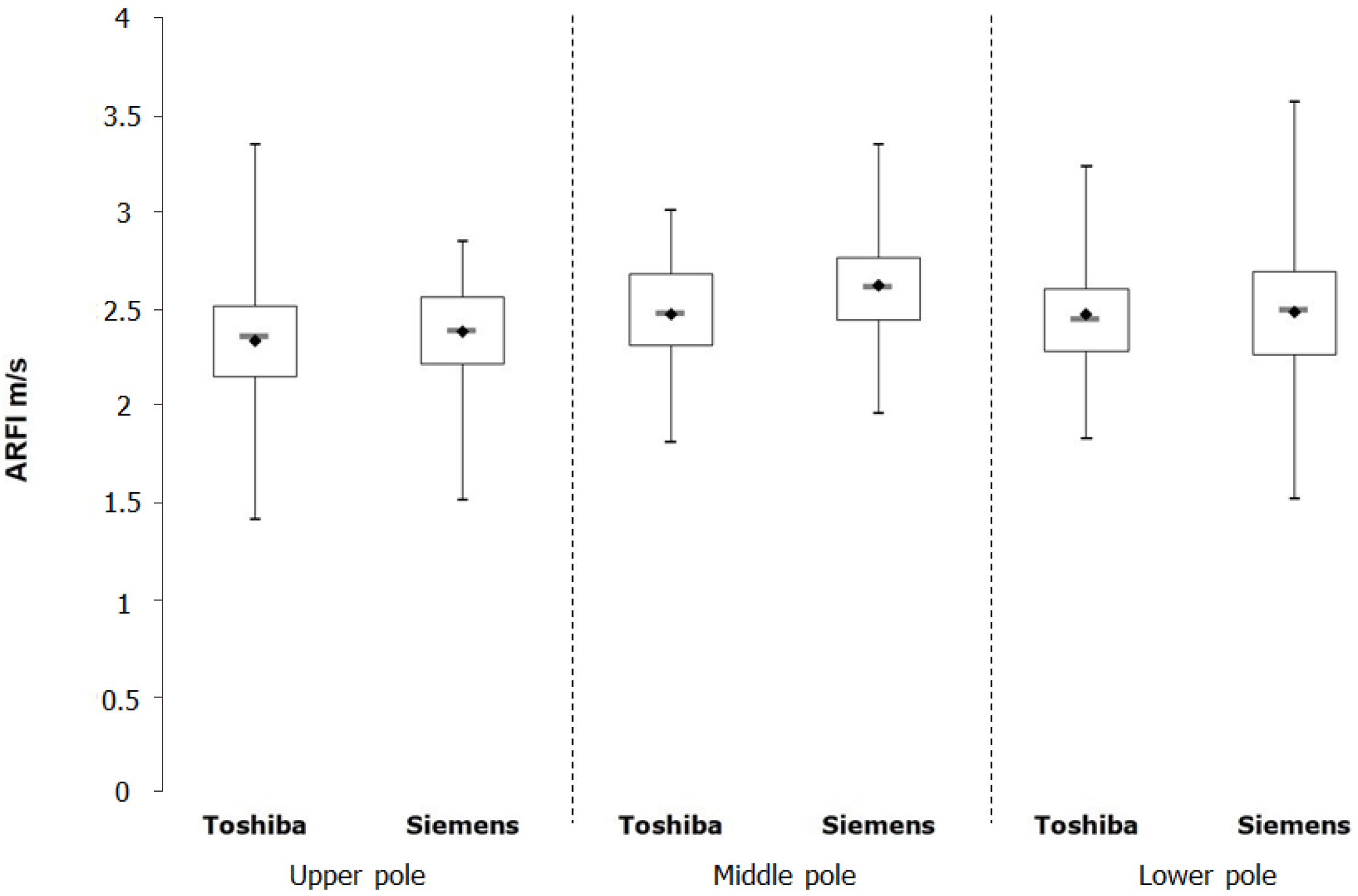
In study arm B, the Siemens device was compared against a newer software version (6.0) of the Toshiba Aplio 500 device. Using the mean values an controlling for age, BMI, and gender, a significant correlation of the shear wave velocities of the two devices was shown for the upper and lower thirds of the spleen (Tables 2 and 4, Figure 3).
In study arm B, no correlation was found between the measured heavy-wave velocities and gender or BMI for both devices tested. In addition, no correlation could be demonstrated for age and shear wave velocity with the Siemens device. Only for the Toshiba Aplio 500 (version 6.0) did we find a significant correlation between age and the measured shear wave velocities, but only for the lower third of the spleen (P < 0.05).
With the help of the subgroup of 44 subjects, we compared the measurements made with the two software versions of the Toshiba Aplio 500 (Tables 1 and 3). All shear wave values obtained with the version 6.0 were significantly lower than those obtained with version 5.0 (P < 0.0001). The mean values differed by 33.0% in the upper third (3.46 m/s vs 2.32 m/s), by 14.0% in the middle third (2.94 m/s vs 2.53 m/s), and by 25.4% in the lower third (3.38 m/s vs 2.52 m/s).
This study is the first to compare four ARFI-based ultrasound elastography methods, two pSWE techniques and two 2D-SWE techniques, from different manufacturers in healthy volunteers taking into account the anatomical location of the measurement of the spleen. Our results show that the anatomical position must be taken into account for splenic elastography. The best results were obtained with the lower pole of the spleen. Furthermore, when interpreting the results using different elastography techniques, attention must be paid to possible limitations in device compatibility. The absolute values of the shear wave elastography measurements of the spleen are not transferable between different manufacturers or models.
In previous studies of the spleen, the measurements were performed at undefined areas or different splenic poles (upper, middle, lower third)[22-26]. Giuffrè et al[33] preferably investigated the lower pole, Albayrak et al[21] performed shear wave elastography of the middle third of the spleen, and Karlas et al[34] performed measurements in an insufficiently defined area between the middle and lower thirds of the spleen. Our results show that the lower third of the spleen is the best anatomical measurement position due to good visibility, as shown by other research groups[26,35-37]. Our results also confirm the recommendations of the EFSUMB to perform elastography on the lower third of the spleen[16]. The upper third does not seem to be suitable for measurements because its anatomical position often makes it difficult or impossible to see by inspiration, as it is partly overlapped by the lung or intestinal segments and located far away from the transducer. Our results confirm that readings should not be assumed to be transferable from one anatomic region of the spleen to another. Whether this is due to the tissue itself or to the examination conditions, such as poor visibility of the upper third, is currently not clear. A previous study reported that the measurement differences between devices and investigators can be up to 15%[38]. In a recent study patients with chronic hepatitis C virus infection show a good agreement of p-SWE and 2D-SWE in patients with F2-F4 fibrosis[39]. Since only healthy subjects were examined in our collective, these results cannot simply be transferred to the situation in patients with chronic hepatitis C and to the spleen[39]. In addition, our results show that without considering the anatomic site of measurement for splenic elastography, reliable measurement results cannot be obtained, regardless of the method used. Again, our results confirm the recommendations of medical societies that the absolute shear wave values are not comparable between different systems and manufacturers[5].
We could not demonstrate any correlation between age and the measured shear wave velocities. This finding is in accordance with the results of recent publications that could not demonstrate any influence of age on the measured shear wave velocities regardless of the shear wave elastography technique used[20,21,33,40,41]. However, an age-related correlation was previously demonstrated in children and adolescents younger than 18 years[42,43]. Independent of the shear wave technique, our study showed a contradictory picture regarding the influence of gender on the measured heavy wave velocities. For both the Siemens device (p-SWE) and the GE device (2D-SWE), gender-specific shear wave velocities were detected. This was not possible for the Philips device (p-SWE). Most of the available studies could not prove any gender-specific influence on the shear wave velocities[21,35,42,44]. A study of healthy children and adolescents concluded that gender influences elastography at the spleen[43]. The influence of BMI on spleen shear wave velocity was not clear according to other research groups[21,33,44]. The influence of abdominal wall thickness on shear wave velocities has not yet been clarified[17,19,20,26] and this parameter was not assessed in our study. Future studies investigating BMI and abdominal wall thickness as influencing factors seem to be necessary.
A limitation of our study is that the defined exclusion criteria were only inquired about anamnestically, and advanced or still undiagnosed diseases could only be excluded by abdominal ultrasonography. Here, in contrast to other studies, no laboratory parameters were determined[22,24]. Also no information on unsuccessful mesaurements was collected during the study. However, due to the predominantly healthy young and slim probands, a low number of unsuccessful measurements can be assumed[45]. Histological examination could not be performed either, as this was ethically unacceptable in young healthy subjects. Compared to current studies and recommendations regarding liver elastography, the low number of measurements in our study is a major limitation. At each position, one measurement was performed with 2D-SWE and five measurements with pSWE. The EFSUMB currently recommends three to five measurements for 2D-SWE in order to obtain good measurements[40]. Karlas et al[34] also recommend between eight and ten measurements for pSWE of the spleen in order to obtain the most accurate shear wave velocities.
In conclusion, the absolute values of the shear wave elastography measurements of the spleen and the two different elastography methods are not comparable between different manufacturers or models. Further studies are needed to confirm the present study results.
Measurement of shear wave velocity in the spleen has been increasingly used in prognostic assessment of esophageal varices and as a marker of portal hypertension. Few systematic comparative studies of the different methods of physical elastography of the spleen are currently available.
Currently, whether the different elastography methods and shear wave measurements with different ultrasonic devices provide comparable results have not been clarified.
The objective of the study was to compare point shear wave and two-dimensional elastography of the spleen considering the anatomical location (upper, hilar, and lower pole).
As part of a prospective clinical study, healthy volunteers were examined for splenic elasticity using four different ultrasound devices between May 2015 and April 2017. The devices used for point shear wave elastography were from Siemens (S 3000) and Philips (Epiq 7), and those used for two-dimensional shear wave elastography were from GE (Logiq E9) and Toshiba (Aplio 500). In addition, two different software versions (5.0 and 6.0) were evaluated for the Toshiba ultrasound device (Aplio 500). The study consisted of three arms: A, B, and C.
In study arm A, 200 subjects were evaluated (78 males and 122 females, mean age 27.9 ± 8.1 years). In study arm B, 113 subjects were evaluated (38 men and 75 women, mean age 26.0 ± 6.3 years). In study arm C, 44 subjects were enrolled. A significant correlation of the shear wave velocities at the upper third of the spleen (r = 0.33088, P < 0.0001) was demonstrated only for the Philips Epiq 7 device compared to the Siemens Acuson S 3000. In comparisons of the other ultrasound devices (GE, Siemens, Toshiba), no comparable results could be obtained for any anatomical position of the spleen. The influencing factors age, gender, and body mass index did not show a clear correlation with the measured shear wave velocities.
The absolute values of the shear wave elastography measurements of the spleen and the two different elastography methods are not comparable between different manufacturers or models.
However, absolute values of splenic shear wave elastography measurements are not transferable between manufacturers or models.
Members of the Elastography Study Group: Hadeel Gamal El-Deen Abd El-Moniem, Gräter Tilmann, Hesse Julian, Klimesch Benjamin, Maaß Marie, Schall Katrin.
| 1. | Li J, Chen M, Cao CL, Zhou LQ, Li SG, Ge ZK, Zhang WH, Xu JW, Cui XW, Dietrich CF. Diagnostic Performance of Acoustic Radiation Force Impulse Elastography for the Differentiation of Benign and Malignant Superficial Lymph Nodes: A Meta-analysis. J Ultrasound Med. 2020;39:213-222. [RCA] [PubMed] [DOI] [Full Text] [Cited by in Crossref: 8] [Cited by in RCA: 8] [Article Influence: 1.3] [Reference Citation Analysis (0)] |
| 2. | Dietrich CF, Hocke M. Elastography of the Pancreas, Current View. Clin Endosc. 2019;52:533-540. [RCA] [PubMed] [DOI] [Full Text] [Full Text (PDF)] [Cited by in Crossref: 36] [Cited by in RCA: 32] [Article Influence: 4.6] [Reference Citation Analysis (0)] |
| 3. | Wee TC, Simon NG. Ultrasound elastography for the evaluation of peripheral nerves: A systematic review. Muscle Nerve. 2019;60:501-512. [RCA] [PubMed] [DOI] [Full Text] [Cited by in Crossref: 42] [Cited by in RCA: 67] [Article Influence: 9.6] [Reference Citation Analysis (0)] |
| 4. | Cornelson SM, Ruff AN, Perillat M, Kettner NW. Sonoelastography of the trunk and lower extremity muscles in a case of Duchenne muscular dystrophy. J Ultrasound. 2019 epub ahead of print. [RCA] [PubMed] [DOI] [Full Text] [Cited by in Crossref: 2] [Cited by in RCA: 5] [Article Influence: 0.7] [Reference Citation Analysis (0)] |
| 5. | Dietrich CF, Bamber J, Berzigotti A, Bota S, Cantisani V, Castera L, Cosgrove D, Ferraioli G, Friedrich-Rust M, Gilja OH, Goertz RS, Karlas T, de Knegt R, de Ledinghen V, Piscaglia F, Procopet B, Saftoiu A, Sidhu PS, Sporea I, Thiele M. EFSUMB Guidelines and Recommendations on the Clinical Use of Liver Ultrasound Elastography, Update 2017 (Long Version). Ultraschall Med. 2017;38:e16-e47. [RCA] [PubMed] [DOI] [Full Text] [Cited by in Crossref: 601] [Cited by in RCA: 607] [Article Influence: 67.4] [Reference Citation Analysis (0)] |
| 6. | Kim DW, Park C, Yoon HM, Jung AY, Lee JS, Jung SC, Cho YA. Technical performance of shear wave elastography for measuring liver stiffness in pediatric and adolescent patients: a systematic review and meta-analysis. Eur Radiol. 2019;29:2560-2572. [RCA] [PubMed] [DOI] [Full Text] [Cited by in Crossref: 9] [Cited by in RCA: 14] [Article Influence: 2.0] [Reference Citation Analysis (0)] |
| 7. | Bota S, Herkner H, Sporea I, Salzl P, Sirli R, Neghina AM, Peck-Radosavljevic M. Meta-analysis: ARFI elastography vs transient elastography for the evaluation of liver fibrosis. Liver Int. 2013;33:1138-1147. [RCA] [PubMed] [DOI] [Full Text] [Cited by in Crossref: 351] [Cited by in RCA: 332] [Article Influence: 25.5] [Reference Citation Analysis (0)] |
| 8. | Sporea I, Bota S, Jurchis A, Sirli R, Grădinaru-Tascău O, Popescu A, Ratiu I, Szilaski M. Acoustic radiation force impulse and supersonic shear imaging vs transient elastography for liver fibrosis assessment. Ultrasound Med Biol. 2013;39:1933-1941. [RCA] [PubMed] [DOI] [Full Text] [Cited by in Crossref: 72] [Cited by in RCA: 65] [Article Influence: 5.0] [Reference Citation Analysis (0)] |
| 9. | Mulazzani L, Salvatore V, Ravaioli F, Allegretti G, Matassoni F, Granata R, Ferrarini A, Stefanescu H, Piscaglia F. Point shear wave ultrasound elastography with Esaote compared to real-time 2D shear wave elastography with supersonic imagine for the quantification of liver stiffness. J Ultrasound. 2017;20:213-225. [RCA] [PubMed] [DOI] [Full Text] [Cited by in Crossref: 22] [Cited by in RCA: 24] [Article Influence: 2.7] [Reference Citation Analysis (0)] |
| 10. | Gress VS, Glawion EN, Schmidberger J, Kratzer W. Comparison of Liver Shear Wave Elastography Measurements using Siemens Acuson S3000, GE LOGIQ E9, Philips EPIQ7 and Toshiba Aplio 500 (Software Versions 5.0 and 6.0) in Healthy Volunteers. Ultraschall Med. 2019;40:504-512. [RCA] [PubMed] [DOI] [Full Text] [Cited by in Crossref: 9] [Cited by in RCA: 14] [Article Influence: 2.0] [Reference Citation Analysis (0)] |
| 11. | Ferraioli G, De Silvestri A, Lissandrin R, Maiocchi L, Tinelli C, Filice C, Barr RG. Evaluation of Inter-System Variability in Liver Stiffness Measurements. Ultraschall Med. 2019;40:64-75. [RCA] [PubMed] [DOI] [Full Text] [Cited by in Crossref: 56] [Cited by in RCA: 56] [Article Influence: 8.0] [Reference Citation Analysis (0)] |
| 12. | Mjelle AB, Mulabecirovic A, Havre RF, Rosendahl K, Juliusson PB, Olafsdottir E, Gilja OH, Vesterhus M. Normal Liver Stiffness Values in Children: A Comparison of Three Different Elastography Methods. J Pediatr Gastroenterol Nutr. 2019;68:706-712. [RCA] [PubMed] [DOI] [Full Text] [Full Text (PDF)] [Cited by in Crossref: 30] [Cited by in RCA: 49] [Article Influence: 7.0] [Reference Citation Analysis (0)] |
| 13. | Mazur R, Celmer M, Silicki J, Hołownia D, Pozowski P, Międzybrodzki K. Clinical applications of spleen ultrasound elastography - a review. J Ultrason. 2018;18:37-41. [RCA] [PubMed] [DOI] [Full Text] [Full Text (PDF)] [Cited by in Crossref: 21] [Cited by in RCA: 20] [Article Influence: 2.5] [Reference Citation Analysis (0)] |
| 14. | Gibiino G, Garcovich M, Ainora ME, Zocco MA. Spleen ultrasound elastography: state of the art and future directions - a systematic review. Eur Rev Med Pharmacol Sci. 2019;23:4368-4381. [RCA] [PubMed] [DOI] [Full Text] [Cited by in RCA: 5] [Reference Citation Analysis (0)] |
| 15. | Săftoiu A, Gilja OH, Sidhu PS, Dietrich CF, Cantisani V, Amy D, Bachmann-Nielsen M, Bob F, Bojunga J, Brock M, Calliada F, Clevert DA, Correas JM, D'Onofrio M, Ewertsen C, Farrokh A, Fodor D, Fusaroli P, Havre RF, Hocke M, Ignee A, Jenssen C, Klauser AS, Kollmann C, Radzina M, Ramnarine KV, Sconfienza LM, Solomon C, Sporea I, Ștefănescu H, Tanter M, Vilmann P. The EFSUMB Guidelines and Recommendations for the Clinical Practice of Elastography in Non-Hepatic Applications: Update 2018. Ultraschall Med. 2019;40:425-453. [RCA] [PubMed] [DOI] [Full Text] [Cited by in Crossref: 220] [Cited by in RCA: 214] [Article Influence: 30.6] [Reference Citation Analysis (0)] |
| 16. | Song J, Huang J, Huang H, Liu S, Luo Y. Performance of spleen stiffness measurement in prediction of clinical significant portal hypertension: A meta-analysis. Clin Res Hepatol Gastroenterol. 2018;42:216-226. [RCA] [PubMed] [DOI] [Full Text] [Cited by in Crossref: 37] [Cited by in RCA: 55] [Article Influence: 6.9] [Reference Citation Analysis (0)] |
| 17. | Cassinotto C, Charrie A, Mouries A, Lapuyade B, Hiriart JB, Vergniol J, Gaye D, Hocquelet A, Charbonnier M, Foucher J, Laurent F, Chermak F, Montaudon M, de Ledinghen V. Liver and spleen elastography using supersonic shear imaging for the non-invasive diagnosis of cirrhosis severity and oesophageal varices. Dig Liver Dis. 2015;47:695-701. [RCA] [PubMed] [DOI] [Full Text] [Cited by in Crossref: 57] [Cited by in RCA: 58] [Article Influence: 5.3] [Reference Citation Analysis (0)] |
| 18. | Takuma Y, Nouso K, Morimoto Y, Tomokuni J, Sahara A, Takabatake H, Matsueda K, Yamamoto H. Portal Hypertension in Patients with Liver Cirrhosis: Diagnostic Accuracy of Spleen Stiffness. Radiology. 2016;279:609-619. [RCA] [PubMed] [DOI] [Full Text] [Cited by in Crossref: 94] [Cited by in RCA: 113] [Article Influence: 10.3] [Reference Citation Analysis (0)] |
| 19. | Balakrishnan M, Souza F, Muñoz C, Augustin S, Loo N, Deng Y, Ciarleglio M, Garcia-Tsao G. Liver and Spleen Stiffness Measurements by Point Shear Wave Elastography via Acoustic Radiation Force Impulse: Intraobserver and Interobserver Variability and Predictors of Variability in a US Population. J Ultrasound Med. 2016;35:2373-2380. [RCA] [PubMed] [DOI] [Full Text] [Cited by in Crossref: 31] [Cited by in RCA: 33] [Article Influence: 3.3] [Reference Citation Analysis (0)] |
| 20. | Cho YS, Lim S, Kim Y, Sohn JH, Jeong JY. Spleen Stiffness Measurement Using 2-Dimensional Shear Wave Elastography: The Predictors of Measurability and the Normal Spleen Stiffness Value. J Ultrasound Med. 2019;38:423-431. [RCA] [PubMed] [DOI] [Full Text] [Cited by in Crossref: 19] [Cited by in RCA: 19] [Article Influence: 2.7] [Reference Citation Analysis (0)] |
| 21. | Albayrak E, Server S. The relationship of spleen stiffness value measured by shear wave elastography with age, gender, and spleen size in healthy volunteers. J Med Ultrason (2001). 2019;46:195-199. [RCA] [PubMed] [DOI] [Full Text] [Cited by in Crossref: 12] [Cited by in RCA: 20] [Article Influence: 2.9] [Reference Citation Analysis (1)] |
| 22. | Ferraioli G, Tinelli C, Lissandrin R, Zicchetti M, Bernuzzi S, Salvaneschi L, Filice C; Elastography Study Group. Ultrasound point shear wave elastography assessment of liver and spleen stiffness: effect of training on repeatability of measurements. Eur Radiol. 2014;24:1283-1289. [RCA] [PubMed] [DOI] [Full Text] [Cited by in Crossref: 74] [Cited by in RCA: 67] [Article Influence: 5.6] [Reference Citation Analysis (0)] |
| 23. | Gallotti A, D'Onofrio M, Pozzi Mucelli R. Acoustic Radiation Force Impulse (ARFI) technique in ultrasound with Virtual Touch tissue quantification of the upper abdomen. Radiol Med. 2010;115:889-897. [RCA] [PubMed] [DOI] [Full Text] [Cited by in Crossref: 124] [Cited by in RCA: 133] [Article Influence: 8.3] [Reference Citation Analysis (0)] |
| 24. | Gao J, Ran HT, Ye XP, Zheng YY, Zhang DZ, Wang ZG. The stiffness of the liver and spleen on ARFI Imaging pre and post TIPS placement: a preliminary observation. Clin Imaging. 2012;36:135-141. [RCA] [PubMed] [DOI] [Full Text] [Cited by in Crossref: 34] [Cited by in RCA: 36] [Article Influence: 2.6] [Reference Citation Analysis (0)] |
| 25. | Grgurevic I, Cikara I, Horvat J, Lukic IK, Heinzl R, Banic M, Kujundzic M, Brkljacic B. Noninvasive assessment of liver fibrosis with acoustic radiation force impulse imaging: increased liver and splenic stiffness in patients with liver fibrosis and cirrhosis. Ultraschall Med. 2011;32:160-166. [RCA] [PubMed] [DOI] [Full Text] [Cited by in Crossref: 38] [Cited by in RCA: 38] [Article Influence: 2.5] [Reference Citation Analysis (0)] |
| 26. | Kaminuma C, Tsushima Y, Matsumoto N, Kurabayashi T, Taketomi-Takahashi A, Endo K. Reliable measurement procedure of virtual touch tissue quantification with acoustic radiation force impulse imaging. J Ultrasound Med. 2011;30:745-751. [RCA] [PubMed] [DOI] [Full Text] [Cited by in Crossref: 49] [Cited by in RCA: 42] [Article Influence: 2.8] [Reference Citation Analysis (0)] |
| 27. | Dong Y, Sirli R, Ferraioli G, Sporea I, Chiorean L, Cui X, Fan M, Wang WP, Gilja OH, Sidhu PS, Dietrich CF. Shear wave elastography of the liver - review on normal values. Z Gastroenterol. 2017;55:153-166. [RCA] [PubMed] [DOI] [Full Text] [Cited by in Crossref: 29] [Cited by in RCA: 30] [Article Influence: 3.3] [Reference Citation Analysis (0)] |
| 28. | Galgenmueller S, Jaeger H, Kratzer W, Schmidt SA, Oeztuerk S, Haenle MM, Mason RA, Graeter T. Parameters affecting different acoustic radiation force impulse applications in the diagnosis of fibrotic liver changes. World J Gastroenterol. 2015;21:8425-8432. [RCA] [PubMed] [DOI] [Full Text] [Full Text (PDF)] [Cited by in CrossRef: 6] [Cited by in RCA: 6] [Article Influence: 0.5] [Reference Citation Analysis (0)] |
| 29. | Keller J, Kaltenbach TE, Haenle MM, Oeztuerk S, Graeter T, Mason RA, Seufferlein T, Kratzer W. Comparison of Acoustic Structure Quantification (ASQ), shearwave elastography and histology in patients with diffuse hepatopathies. BMC Med Imaging. 2015;15:58. [RCA] [PubMed] [DOI] [Full Text] [Full Text (PDF)] [Cited by in Crossref: 13] [Cited by in RCA: 16] [Article Influence: 1.5] [Reference Citation Analysis (0)] |
| 30. | General Assembly of the World Medical Association. . World Medical Association Declaration of Helsinki: ethical principles for medical research involving human subjects. J Am Coll Dent. 2014;81:14-18. [PubMed] |
| 31. | Serra C, Grasso V, Conti F, Felicani C, Mazzotta E, Lenzi M, Verucchi G, D'errico A, Andreone P. A New Two-Dimensional Shear Wave Elastography for Noninvasive Assessment of Liver Fibrosis in Healthy Subjects and in Patients with Chronic Liver Disease. Ultraschall Med. 2018;39:432-439. [RCA] [PubMed] [DOI] [Full Text] [Cited by in Crossref: 22] [Cited by in RCA: 16] [Article Influence: 2.0] [Reference Citation Analysis (0)] |
| 32. | Fang C, Konstantatou E, Romanos O, Yusuf GT, Quinlan DJ, Sidhu PS. Reproducibility of 2-Dimensional Shear Wave Elastography Assessment of the Liver: A Direct Comparison With Point Shear Wave Elastography in Healthy Volunteers. J Ultrasound Med. 2017;36:1563-1569. [RCA] [PubMed] [DOI] [Full Text] [Cited by in Crossref: 33] [Cited by in RCA: 35] [Article Influence: 3.9] [Reference Citation Analysis (0)] |
| 33. | Giuffrè M, Macor D, Masutti F, Abazia C, Tinè F, Patti R, Buonocore MR, Colombo A, Visintin A, Campigotto M, Crocè LS. Evaluation of spleen stiffness in healthy volunteers using point shear wave elastography. Ann Hepatol. 2019;18:736-741. [RCA] [PubMed] [DOI] [Full Text] [Cited by in Crossref: 30] [Cited by in RCA: 43] [Article Influence: 7.2] [Reference Citation Analysis (0)] |
| 34. | Karlas T, Lindner F, Tröltzsch M, Keim V. Assessment of spleen stiffness using acoustic radiation force impulse imaging (ARFI): definition of examination standards and impact of breathing maneuvers. Ultraschall Med. 2014;35:38-43. [RCA] [PubMed] [DOI] [Full Text] [Cited by in Crossref: 20] [Cited by in RCA: 23] [Article Influence: 1.9] [Reference Citation Analysis (0)] |
| 35. | Goertz RS, Amann K, Heide R, Bernatik T, Neurath MF, Strobel D. An abdominal and thyroid status with Acoustic Radiation Force Impulse Elastometry--a feasibility study: Acoustic Radiation Force Impulse Elastometry of human organs. Eur J Radiol. 2011;80:e226-e230. [RCA] [PubMed] [DOI] [Full Text] [Cited by in Crossref: 76] [Cited by in RCA: 79] [Article Influence: 4.9] [Reference Citation Analysis (1)] |
| 36. | Mori K, Arai H, Abe T, Takayama H, Toyoda M, Ueno T, Sato K. Spleen stiffness correlates with the presence of ascites but not esophageal varices in chronic hepatitis C patients. Biomed Res Int. 2013;2013:857862. [RCA] [PubMed] [DOI] [Full Text] [Full Text (PDF)] [Cited by in Crossref: 14] [Cited by in RCA: 12] [Article Influence: 0.9] [Reference Citation Analysis (0)] |
| 37. | Procopet B, Berzigotti A, Abraldes JG, Turon F, Hernandez-Gea V, García-Pagán JC, Bosch J. Real-time shear-wave elastography: applicability, reliability and accuracy for clinically significant portal hypertension. J Hepatol. 2015;62:1068-1075. [RCA] [PubMed] [DOI] [Full Text] [Cited by in Crossref: 178] [Cited by in RCA: 161] [Article Influence: 14.6] [Reference Citation Analysis (0)] |
| 38. | Hall TJ, Milkowski A, Garra B, Carson P, Palmeri M, Nightingale K, Lynch T, Alturki A, Andre M, Audiere S, Bamber J, Barr R, Bercoff J, Bernal M, Brum J, Cohen-Bacrie C, Couade M, Daniels A, DeWall R, Dillman J, Ehman R, Franchi-Abella SF, Fromageau J, Gennisson J, Henry JP, Ivancevich N, Kalin J, Kohn S, Kugel J, Liu N L, Loupas T, Mazernik J, McAleavey S, Miette V, Metz S, Morel BM, Nelson T, Nordberg E, Oudry J, Padwal M, Rouze N, Samir A, Sandrin L, Schaccitti J, Schmitt C, Shamdasani V, Switalski P, Wang M, Wear K. RSNA/QIBA: Shear wave speed as a biomarker for liver fibrosis staging. 2013 IEEE International Ultrasonics Symposium (IUS); 2013 July 21-25; Prague, Czech Republic. [DOI] [Full Text] |
| 39. | Bâldea V, Lupușoru R, Dănilă M, Șirli R, Popescu A, Sporea I. Comparison between the performance of Two-Dimensional and Point Shear Wave elastography for the noninvasive assessment of liver cirrhosis. Ultrasound Med Biol. 2019;45:S119. [DOI] [Full Text] |
| 40. | Arda K, Ciledag N, Aktas E, Aribas BK, Köse K. Quantitative assessment of normal soft-tissue elasticity using shear-wave ultrasound elastography. AJR Am J Roentgenol. 2011;197:532-536. [RCA] [PubMed] [DOI] [Full Text] [Full Text (PDF)] [Cited by in Crossref: 282] [Cited by in RCA: 336] [Article Influence: 25.8] [Reference Citation Analysis (0)] |
| 41. | Pawluś A, Inglot MS, Szymańska K, Kaczorowski K, Markiewicz BD, Kaczorowska A, Gąsiorowski J, Szymczak A, Inglot M, Bladowska J, Zaleska-Dorobisz U. Shear wave elastography of the spleen: evaluation of spleen stiffness in healthy volunteers. Abdom Radiol (NY). 2016;41:2169-2174. [RCA] [PubMed] [DOI] [Full Text] [Full Text (PDF)] [Cited by in Crossref: 26] [Cited by in RCA: 38] [Article Influence: 3.8] [Reference Citation Analysis (1)] |
| 42. | Cañas T, Fontanilla T, Miralles M, Maciá A, Malalana A, Román E. Normal values of spleen stiffness in healthy children assessed by acoustic radiation force impulse imaging (ARFI): comparison between two ultrasound transducers. Pediatr Radiol. 2015;45:1316-1322. [RCA] [PubMed] [DOI] [Full Text] [Cited by in Crossref: 13] [Cited by in RCA: 15] [Article Influence: 1.4] [Reference Citation Analysis (0)] |
| 43. | Lee MJ, Kim MJ, Han KH, Yoon CS. Age-related changes in liver, kidney, and spleen stiffness in healthy children measured with acoustic radiation force impulse imaging. Eur J Radiol. 2013;82:e290-e294. [RCA] [PubMed] [DOI] [Full Text] [Cited by in Crossref: 78] [Cited by in RCA: 90] [Article Influence: 6.9] [Reference Citation Analysis (0)] |
| 44. | Kassym L, Nounou MA, Zhumadilova Z, Dajani AI, Barkibayeva N, Myssayev A, Rakhypbekov T, Abuhammour AM. New combined parameter of liver and splenic stiffness as determined by elastography in healthy volunteers. Saudi J Gastroenterol. 2016;22:324-330. [RCA] [PubMed] [DOI] [Full Text] [Full Text (PDF)] [Cited by in Crossref: 6] [Cited by in RCA: 7] [Article Influence: 0.7] [Reference Citation Analysis (0)] |
| 45. | Petzold G, Hofer J, Ellenrieder V, Neesse A, Kunsch S. Liver Stiffness Measured by 2-Dimensional Shear Wave Elastography: Prospective Evaluation of Healthy Volunteers and Patients With Liver Cirrhosis. J Ultrasound Med. 2019;38:1769-1777. [RCA] [PubMed] [DOI] [Full Text] [Cited by in Crossref: 16] [Cited by in RCA: 18] [Article Influence: 2.6] [Reference Citation Analysis (0)] |
Open-Access: This article is an open-access article that was selected by an in-house editor and fully peer-reviewed by external reviewers. It is distributed in accordance with the Creative Commons Attribution NonCommercial (CC BY-NC 4.0) license, which permits others to distribute, remix, adapt, build upon this work non-commercially, and license their derivative works on different terms, provided the original work is properly cited and the use is non-commercial. See: http://creativecommons.org/Licenses/by-nc/4.0/
Manuscript source: Unsolicited manuscript
Corresponding Author's Membership in Professional Societies: German Society for Ultrasound in Medicine; German Society for Digestive and Metabolic Diseases; Professional Association of German Internists (Berufsverband Deutscher Internisten); Southwest German Society for Internal Medicine (Südwestdeutsche Gesellschaft für Innere Medizin); Paul-Ehrlich-Society (Paul-Ehrlich-Gesellschaft (Arbeitsgemeinschaft Echinokokkose)); Member of the AG Ultrasound of the DGVS (Mitglied der AG Ultraschall der DGVS); and Member of the American Institute of Ultrasound in Medicine.
Specialty type: Gastroenterology and hepatology
Country/Territory of origin: Germany
Peer-review report’s scientific quality classification
Grade A (Excellent): 0
Grade B (Very good): B
Grade C (Good): C
Grade D (Fair): 0
Grade E (Poor): 0
P-Reviewer: Treeprasertsuk S S-Editor: Liu M L-Editor: A P-Editor: Wang LL


