
















©The Author(s) 2017.
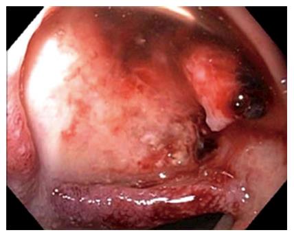
Figure 1 Endoscopic image showing a large ulcer in the superior wall of the bulb with a large visible vessel.
Attempted endoclip placement after epinephrine injection resulted in major bleeding and a loss of endoscopic view and patient was then emergently transferred to interventional radiology.
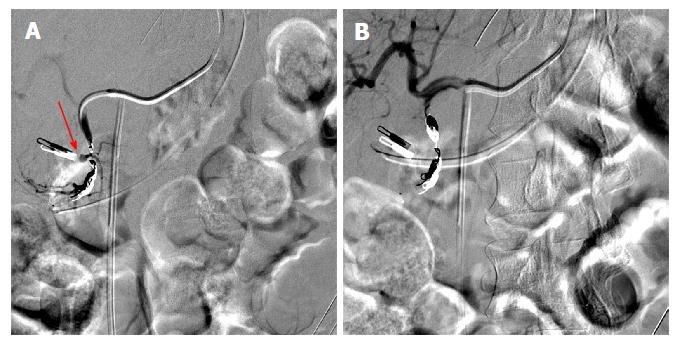
Figure 2 During interventional angiography, selected fluoroscopic images showing a pseudoaneurysm of the gastroduodenal artery (A, red arrow) that was successfully coiled with subsequent hemostasis via the sandwich technique (B).
Previously placed Endoclip is visible and can act as a fluoroscopic marker during angiography.
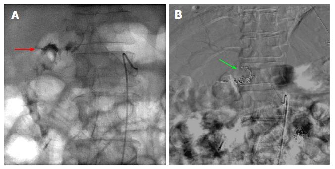
Figure 3 Fluoroscopic images of a case of 2-3 cm bleeding duodenal ulcer that failed endoscopic hemostasis with Endoclip application.
A: Fluoroscopic image following contrast injection to the right gastric artery showing active extravasation into the lumen. The bleeding vessel (red arrow) is identified which is near the endoscopically placed clip; B: Digital subtraction angiography post coiling of the right gastric artery (green arrow).
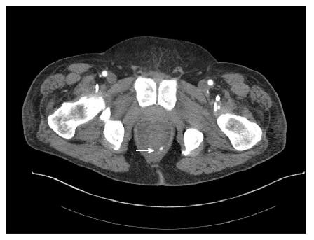
Figure 4 A male patient presented with massive bright red blood per rectum which was unresponsive to transfusions.
A representative computed tomography scan image demonstrates active contrast extravasation in the rectum (white arrow).
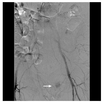
Figure 5 During interventional angiography, contrast extravasation is visualized into the colon via a distal branch artery from the internal iliac artery (white arrow).
The culprit vessel was occluded by spasm or dissection at the ostium with no residual active bleeding. During follow-up lower endoscopy, an endoscopic image showed no active bleeding with discrete large sized clean based ulcers, consistent with ischemic colitis.
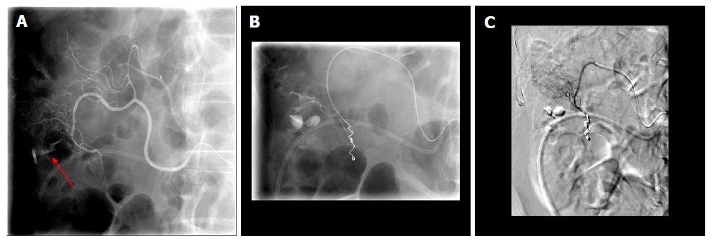
Figure 6 Fluoroscopic images of a case of colon diverticular bleeding that failed endoscopic hemostasis with Endoclip application.
A: Active extravasation at the site of the clip (red arrow); B: Ongoing extravasation superior to the clip after initial coils placed in the inferior branch of the middle colic artery; C: Digital subtraction angiography following additional coils with no extravasation.
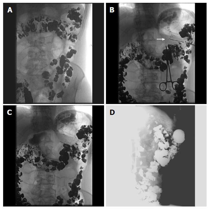
Figure 7 Selected fluoroscopic images demonstrating fluoroscopically guided gastrostomy placement.
A: Contrast seen throughout the transverse colon; B: The first T-tac (white arrow) is deployed following needle insertion into the gastric lumen; C: Two more T-tacs placed; D: Static lateral image of the gastrostomy tube against abdominal wall and not in the colon. The contrast is injected into the tube to demonstrate intraluminal placement.
- Citation: Ray DM, Srinivasan I, Tang SJ, Vilmann AS, Vilmann P, McCowan TC, Patel AM. Complementary roles of interventional radiology and therapeutic endoscopy in gastroenterology. World J Radiol 2017; 9(3): 97-111
- URL: https://www.wjgnet.com/1949-8470/full/v9/i3/97.htm
- DOI: https://dx.doi.org/10.4329/wjr.v9.i3.97