©The Author(s) 2017.
World J Radiol. Oct 28, 2017; 9(10): 400-404
Published online Oct 28, 2017. doi: 10.4329/wjr.v9.i10.400
Published online Oct 28, 2017. doi: 10.4329/wjr.v9.i10.400
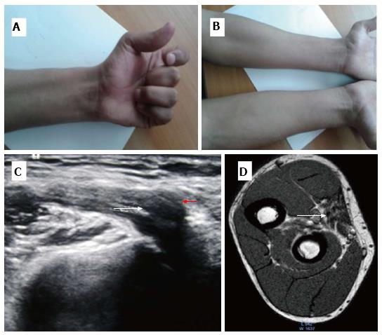
Figure 1 Images of first patient with intraosseous median nerve entrapment.
A: Clinical Photograph showing inability to close fist completely with partial flexion of the index finger; B: Atrophy of anterior compartment muscles of left forearm; C: Sonogram shows thickened and hypoechoic median nerve (arrow), coursing posteriorly through the fractured bone; D: SE T1 W MR axial image shows atrophy of anterior compartment muscles of forearm with fatty infiltration s/o chronic denervation changes (arrow).
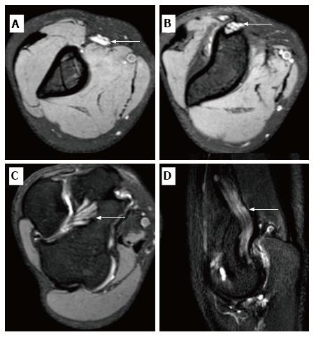
Figure 2 Images of second patient with intraosseous median nerve entrapment.
A-C: 3D GRE T2W images showing markedly thickened and hyperintense entrapped median nerve (arrow), coursing posteriorly through the fractured medial epicondyle; D: STIR Coronal image showing markedly thickened and hyperintense median nerve (arrow).
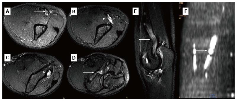
Figure 3 Images of third patient with intraosseous median nerve entrapment.
A-D: SE T2W FS axial MR image shows markedly thickened and hyperintense nerve coursing through the fractured medial epicondyle; E: STIR coronal MR image shows entrapped median nerve coursing posteriorly; F: DWIBS coronal reformat highlights the abnormal median nerve.
- Citation: Aggarwal A, Jana M, Kumar V, Srivastava DN, Garg K. MR neurography in intraosseous median nerve entrapment. World J Radiol 2017; 9(10): 400-404
- URL: https://www.wjgnet.com/1949-8470/full/v9/i10/400.htm
- DOI: https://dx.doi.org/10.4329/wjr.v9.i10.400


