Copyright
©The Author(s) 2016.
World J Radiol. Jul 28, 2016; 8(7): 656-667
Published online Jul 28, 2016. doi: 10.4329/wjr.v8.i7.656
Published online Jul 28, 2016. doi: 10.4329/wjr.v8.i7.656
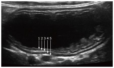
Figure 1 Normal small bowel.
Ultrasound image of small bowel obtained after ingestion of water, using high-resolution linear probe. Five wall layers include: 1-mucosal interface with lumen (hyperechoic), 2-mucosa (hypoechoic), 3-submucosa (hyperechoic), 4-muscularis (hypoechoic), and 5-serosa (hyperechoic).
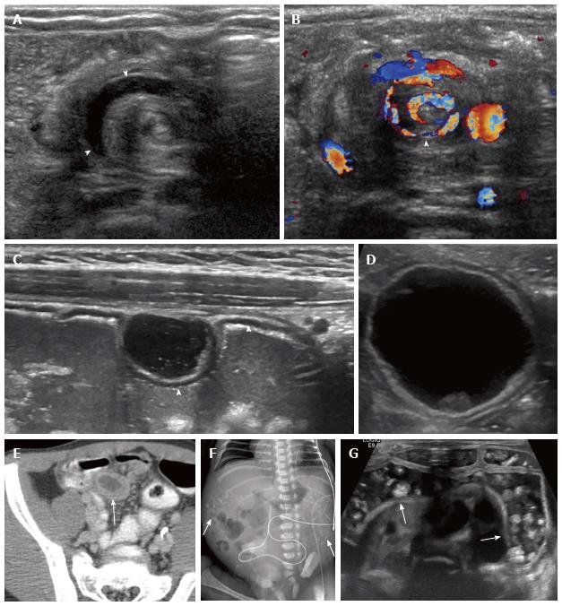
Figure 2 Congenital bowel abnormalities.
A, B: Malrotation. Unsuspected finding in an 8-wk-old vomiting infant being evaluated for pyloric stenosis. Transverse midline images show alternating rings of high and low echogenicity with “whirlpool” sign on grayscale (A) and color Doppler (B) images (arrowheads); C: Gastric duplication cyst. Five-year-old girl with enteric duplication cyst near the gastroesophageal junction detected prenatally (not shown). A second cyst was noted incidentally in the anterior wall of the stomach on subsequent imaging. The cyst demonstrates bowel signature (2 layers), and shares its hypoechoic, muscularis propria layer with the anterior gastric wall (arrowheads); D, E: Meckel diverticulum. Four-year-old with abdominal pain. Ultrasound shows a cyst with bowel signature (D). Computed tomography abdomen is shown for correlation (E, arrow); F, G: Rectourinary fistula with enteroliths. Newborn with abdominal calcifications on radiograph (F, arrows); confirmed to be enteroliths on ultrasound (G, arrows). The fistula was later confirmed with contrast enema (not shown).
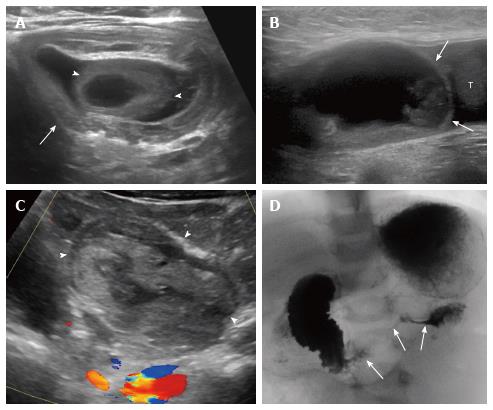
Figure 3 Acquired bowel disorders.
A: Ileocolic intussusception with Meckel diverticulum as lead point. Six-month-old with small bowel obstruction on radiograph (not shown) and intussusception (arrow) demonstrated on ultrasound with lead point (arrowheads); B: Incarcerated inguinal hernia. Two-year-old boy with abdominal pain and left groin mass. Sagittal image of the left inguinal region show a cystic structure that did not clearly communicate with abdominal bowel loops (B, arrows; T = testicle). Testicular edema was also noted (not shown); C, D: Duodenal hematoma secondary to child abuse. One-year-old with abdominal pain and distension. Sagittal midline ultrasound image shows a complex mass in the expected location of the duodenum (C, arrowheads). Upper gastrointestinal series confirmed duodenal narrowing (D, arrows). Abuse was later confirmed.
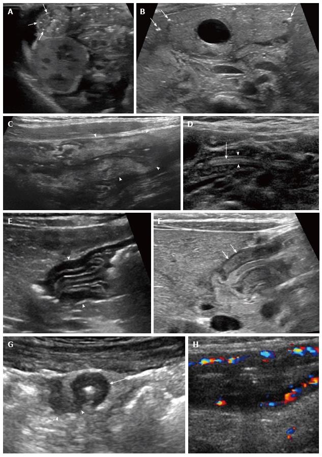
Figure 4 Infectious and inflammatory bowel disorders.
A, B: Necrotizing enterocolitis. Targeted ultrasound of the abdomen in a premature infant shows bowel wall thickening and echogenic intramural air (A, arrows). This corresponded to an area of pneumatosis on recent radiograph (not shown). Transverse image of the liver show punctate, echogenic foci in the liver periphery, consistent with portal venous gas; the foci of air are too small to cause posterior artifact (B, arrows); C: Campylobacter enterocolitis. Ten-year-old with fever and abdominal pain with suspected appendicitis. Sagittal right lower quadrant ultrasound image shows mural thickening and increased echogenicity in the cecum and ascending colon (arrowheads). Stool cultures confirmed the diagnosis; D: Ascariasis. Two-year-old boy from Africa with abdominal pain. Ultrasound of the small bowel shows a mobile, hypoechoic, tubular structure with echogenic walls (arrowheads) and central linear echogenicity (arrow). Worms were later identified in the stool; E, F: Allergic (eosinophilic) gastritis. Ultrasound of the stomach in a 3-mo-old infant with persistent vomiting shows mural thickening in the antrum with prominent mucosal and submucosal layers (E, arrowheads). Endoscopy confirmed the diagnosis. Ultrasound of child with pyloric stenosis (F), for comparison, shows thickening primarily of the muscularis layer (arrows); G, H: Crohn disease. Transverse image of the right lower quadrant in a 15-year-old girl with longstanding Crohn disease shows a thick-walled ileum in cross section (arrow) with a fistula extending posterolaterally (arrowheads), confirmed with MRI (not shown) (G, arrows). Color Doppler ultrasound image in another patient with Crohn disease demonstrates mural thickening and hyperemia of the inflamed terminal ileum (H).
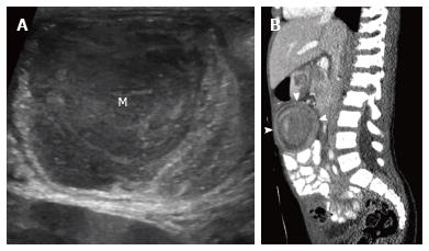
Figure 5 Burkitt lymphoma.
Eight-year-old boy presented with weight loss and abdominal pain. Abdominal ultrasound showed ileocolic intussusception with soft tissue mass (M) as a lead point (A). Bilateral renal masses were also present (not shown). Sagittal reformatted computed tomography image shows ileocolic intussusception (B, arrowheads). Diagnosis was confirmed with biopsy.
Figure 6 Henoch Schonlein purpura.
Seven-year-old boy with purpuric rash and abdominal pain. Ultrasound image with color Doppler shows thick walled and hyperemic small bowel loops (A, arrowheads) and small bowel-small bowel intussusception (A, arrow). Computed tomography shows stratified enhancement of thick walled small bowel with submucosal edema (B, arrowhead).
- Citation: Gale HI, Gee MS, Westra SJ, Nimkin K. Abdominal ultrasonography of the pediatric gastrointestinal tract. World J Radiol 2016; 8(7): 656-667
- URL: https://www.wjgnet.com/1949-8470/full/v8/i7/656.htm
- DOI: https://dx.doi.org/10.4329/wjr.v8.i7.656


