Copyright
©The Author(s) 2016.
World J Radiol. May 28, 2016; 8(5): 501-505
Published online May 28, 2016. doi: 10.4329/wjr.v8.i5.501
Published online May 28, 2016. doi: 10.4329/wjr.v8.i5.501
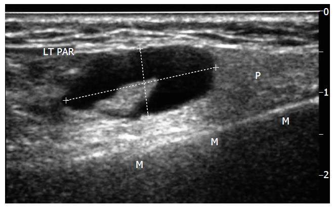
Figure 1 Longitudinal sonogram through left parotid gland (P) superficial lobe, mandible (M).
Demonstrates an atypical node, callipers-eccentric hilum displaced and heterogeneous cortex. LT PAR: Left Parotid.
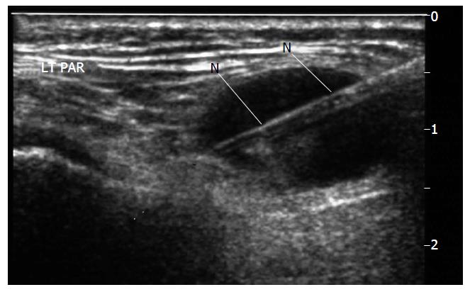
Figure 2 Biopsy undertaken with spring loaded biopsy automated device, 22 mm throw, 18G needle (N).
Note needle deployed to traverse but not exit the lesion. LT PAR: Left Parotid.
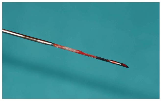
Figure 3 Image of biopsy specimen taken in Figure 2.
18G-needle in biopsy tray with overlying needle overlying sheath retracted.
Figure 4 Low power image, H and E stain, of biopsy samples obtained in this case to give an idea of what material is available to the pathologist for reporting and immunohistochemistry - final diagnosis in this case was B cell non-hodgkins Lymphoma.
- Citation: Haldar S, Sinnott JD, Tekeli KM, Turner SS, Howlett DC. Biopsy of parotid masses: Review of current techniques. World J Radiol 2016; 8(5): 501-505
- URL: https://www.wjgnet.com/1949-8470/full/v8/i5/501.htm
- DOI: https://dx.doi.org/10.4329/wjr.v8.i5.501


