Copyright
©The Author(s) 2016.
World J Radiol. May 28, 2016; 8(5): 484-500
Published online May 28, 2016. doi: 10.4329/wjr.v8.i5.484
Published online May 28, 2016. doi: 10.4329/wjr.v8.i5.484
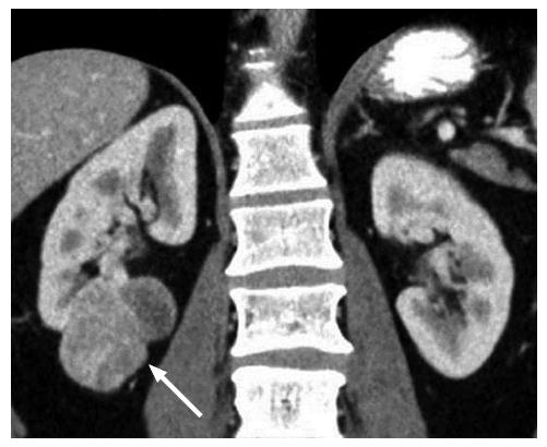
Figure 1 A 45-year-old male with a pathologically proven clear cell renal cell carcinoma in the right kidney on a coronal contrast-enhanced computed tomography image.
The exophytic tumor (arrow) has a heterogeneous solid and cystic internal consistency.
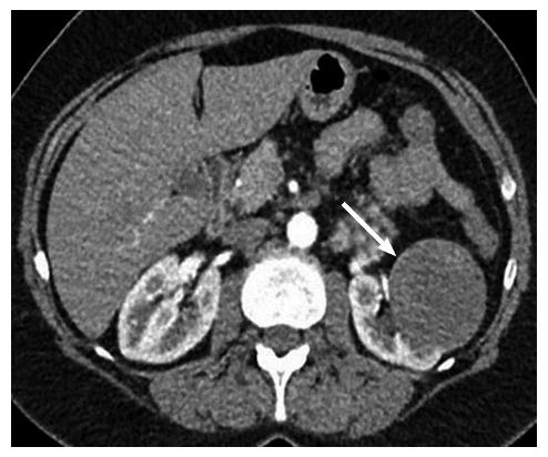
Figure 2 A 47-year-old female with a pathologically proven papillary renal cell carcinoma in the left kidney on an axial contrast-enhanced computed tomography image.
The well-circumscribed hypovascular exophytic tumor (arrow) has a homogeneous solid internal consistency.
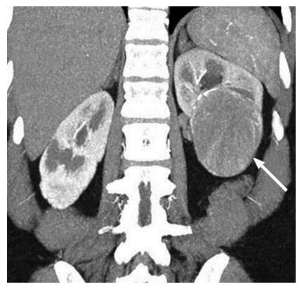
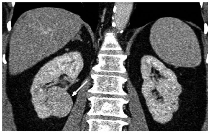
Figure 3 A 53-year-old female with a pathologically proven chromophobe renal cell carcinoma in the left kidney on a coronal contrast-enhanced computed tomography image.
The well-circumscribed tumor (arrow) shows a homogeneous solid consistency and peripheral internal tumor vessels.
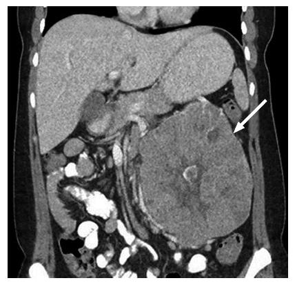
Figure 4 A 36-year-old female with a pathologically proven chromophobe renal cell carcinoma in the left kidney on a coronal contrast-enhanced computed tomography image.
The large well-circumscribed solid tumor (arrow) shows a hypoattenuating central stellate scar and internal calcification.
Figure 5 A 61-year-old female with a pathologically proven clear cell renal cell carcinoma in the right kidney.
A: Axial in-phase T1-weighted magnetic resonance imaging; B: Axial opposed-phase T1-weighted magnetic resonance image; C: Axial fat-only magnetic resonance image from a two-point Dixon reconstruction which displays the difference between echos from A and B. The tumor (arrow) shows high signal on C due to the presence of microscopic fat. Incidentally, the liver also shows high signal on C due to hepatic steatosis.
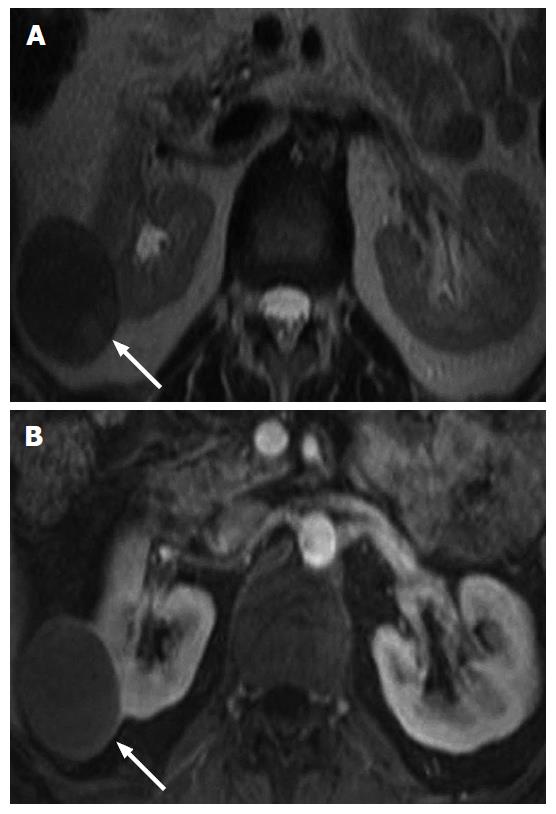
Figure 6 A 42-year-old male with a pathologically proven papillary renal cell carcinoma in the right kidney.
A: On an axial T2-weighted magnetic resonance image. The well-circumscribed exophytic solid tumor (arrow) shows relatively homogeneous low T2 signal intensity; B: On an axial contrast-enhanced T1-weighted magnetic resonance image during the corticomedullary phase. The tumor (arrow) is homogeneously hypovascular compared to the adjacent renal cortex, except for mild enhancement of the renal capsule.
Figure 7 A 61-year-old female with a clear cell renal cell carcinoma in the right kidney.
A: On an axial T2-weighted magnetic resonance image. The solid tumor (arrow) shows a heterogeneous high T2 signal intensity; B: On an axial contrast-enhanced T1-weighted magnetic resonance image during the corticomedullary phase. The solid tumor (arrow) shows heterogeneous hypervascularity, of a similar degree to that of the adjacent normal renal cortex.
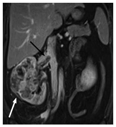
Figure 8 A 42-year-old female with a pathologically proven clear cell renal cell carcinoma in the right kidney on a coronal contrast-enhanced T1-weighted magnetic resonance image.
The ill-marginated tumor (white arrow) involves the whole of the kidney and shows extension into the right renal vein (black arrow) and slight protrusion into the inferior vena cava.
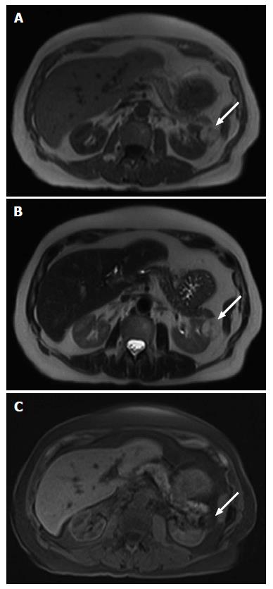
Figure 9 A 81-year-old male with a macroscopic fat containing renal angiomyolipoma.
A: On an axial T1-weighted magnetic resonance image. The ovoid lesion (arrow) in the left kidney shows uniform high T1 signal intensity; B: On an axial T2-weighted magnetic resonance image. The ovoid lesion (arrow) in the left kidney shows uniform high T2 signal intensity; C: On an axial fat suppressed T1-weighted magnetic resonance image. The ovoid lesion (arrow) in the left kidney which previously demonstrated uniform high T1 and T2 signal intensities now shows uniform signal loss.
Figure 10 A 59-year-old female with a pathologically proven oncocytoma in the lower pole of the right kidney on a coronal contrast-enhanced computed tomography image.
The well-circumscribed tumor (arrow) shows a homogeneous solid consistency.
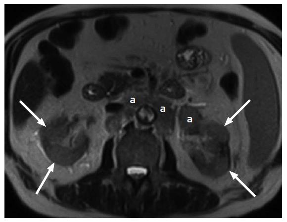
Figure 11 A 62-year-old male with pathologically proven B cell lymphoma on an axial T2-weighted image.
Multifocal bilateral poorly-defined masses (arrows) of intermediate to high T2 signal intensity in the kidneys are due to secondary renal lymphoma. In addition, there is lymphomatous involvement of enlarged retroperitoneal lymph nodes (a) in the para-aortic region.
- Citation: Low G, Huang G, Fu W, Moloo Z, Girgis S. Review of renal cell carcinoma and its common subtypes in radiology. World J Radiol 2016; 8(5): 484-500
- URL: https://www.wjgnet.com/1949-8470/full/v8/i5/484.htm
- DOI: https://dx.doi.org/10.4329/wjr.v8.i5.484


