Copyright
©The Author(s) 2016.
World J Radiol. Apr 28, 2016; 8(4): 428-433
Published online Apr 28, 2016. doi: 10.4329/wjr.v8.i4.428
Published online Apr 28, 2016. doi: 10.4329/wjr.v8.i4.428
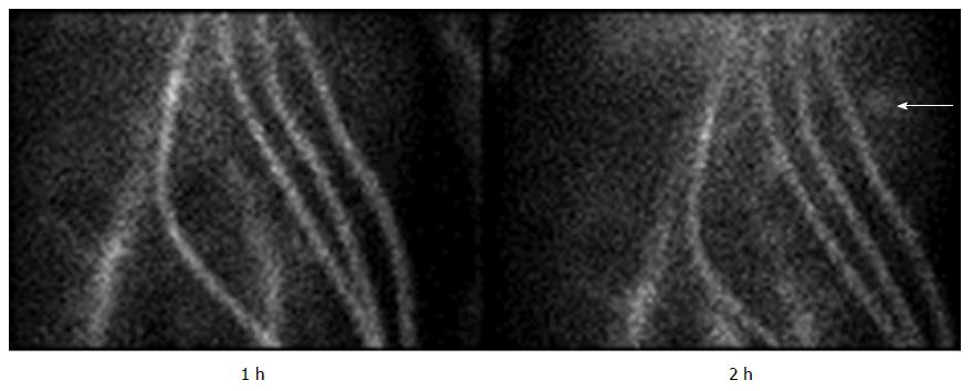
Figure 1 Gastrointestinal bleeding scan showing pooling of blood in the left upper quadrant.
The white arrow indicates the area of bleeding. Note the signal arising from the four cannulae of the continuous-flow biventricular assist devices, as well as iliac vessels, indicating persistent presence of radiolabeled blood in the circulation.
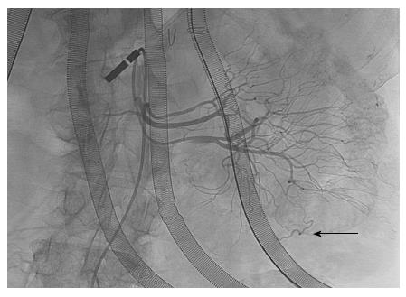
Figure 2 Selective angiogram of proximal jejunal branches of the superior mesenteric artery.
The black arrow indicates the early filling vein, indicative of the presence of a vascular malformation.
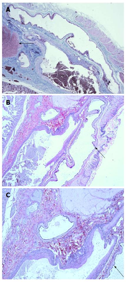
Figure 3 Vascular malformation.
A: Trichrome staining (10 × magnification) of the jejunum showing dilated artery and vein with attenuated muscularis layer (muscularis indicated by arrow); B: Elastic staining (20 ×) showing dark inner lining of vessel indicating presence of internal elastic lamina of artery (black arrow), contrasted with lack of elastic lamina in adjacent vein. This is also shown at 40 × magnification in C.
- Citation: Mirasol RV, Tholany JJ, Reddy H, Fyfe-Kirschner BS, Cheng CL, Moubarak IF, Nosher JL. Gastrointestinal bleeding in a patient with a continuous-flow biventricular assist device. World J Radiol 2016; 8(4): 428-433
- URL: https://www.wjgnet.com/1949-8470/full/v8/i4/428.htm
- DOI: https://dx.doi.org/10.4329/wjr.v8.i4.428


