Copyright
©The Author(s) 2016.
World J Radiol. Apr 28, 2016; 8(4): 342-354
Published online Apr 28, 2016. doi: 10.4329/wjr.v8.i4.342
Published online Apr 28, 2016. doi: 10.4329/wjr.v8.i4.342
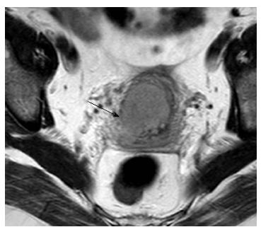
Figure 1 Cervical cancer, stage IIB.
Axial oblique T2-weighted image shows focal disruption of the low signal cervical stroma on the right (arrow), a sign of parametrial invasion.
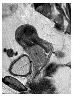
Figure 2 Cervical cancer, stage IIA.
Sagittal T2-weighted image shows a large cervical mass (asterisk) invading the anterior vaginal wall (long arrow). Preservation of the low signal intensity of the posterior vaginal fornix (small arrow) excludes invasion of posterior wall.
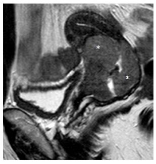
Figure 3 Cervical cancer, stage IB2.
Sagittal T2-weighted image shows a bulky cervical mass (asterisk) with endocervical growth close to internal cervical os.
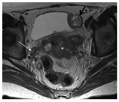
Figure 4 Cervical cancer, stage IVB.
Axial T2-weighted image demonstrates a large cervical mass (asterisk) extending to the pelvic sidewall, on the right (white arrow). A heterogeneous mass consistent with a peritoneal implant is also seen abutting the left side of the bladder (black arrow).
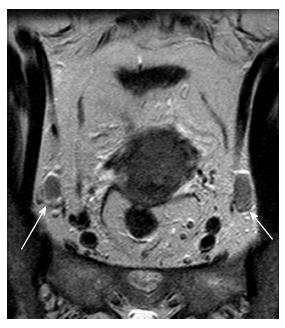
Figure 5 Cervical cancer, stage IIB.
Coronal oblique T2-weighted image demonstrates the presence of enlarged internal obturator lymph nodes, bilaterally (arrows).
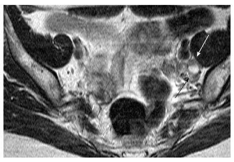
Figure 6 Cervical cancer, stage IB2.
Axial T2-weighted image shows an enlarged left internal obturator node with central necrosis (arrows). Caution should be taken to differentiate from normal ovary.
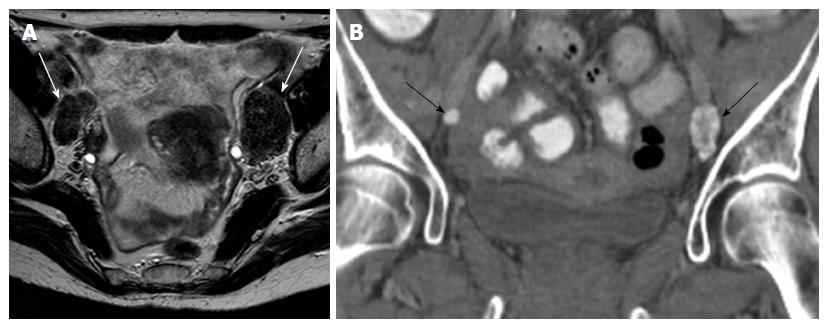
Figure 7 Cervical cancer, stage IIB.
A: Axial T2-weighted image shows bilateral enlarged external iliac lymph nodes with very low signal intensity (white arrows); B: Coronal reconstruction of computed tomography of the pelvis, confirms the presence of calcifications within the nodes (arrows). Calcification within metastatic nodes from cervical cancer is an unusual finding.
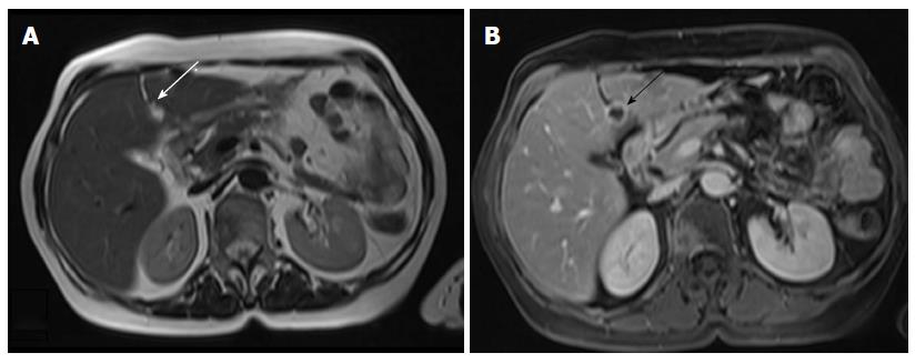
Figure 8 Cervical cancer, stage IVB.
A: Axial T2-weighted image shows a hyperintense lesion within the falciform ligament (arrow); B: Corresponding axial T1-weighted image after intravenous injection of paramagnetic contrast shows the necrotic nature of the lesion (arrow). Cavitating metastatic lesions are usually indicative of cervical carcinomas of squamous cell origin.
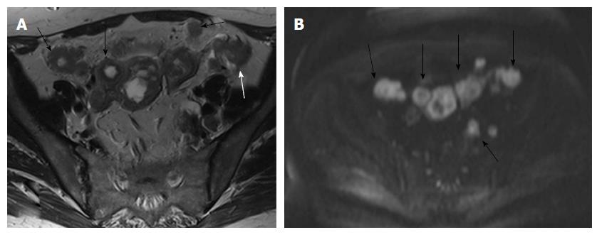
Figure 9 Cervical cancer, stage IVB.
A: Axial T2-weighted image clearly demonstrate multiple tumorous peritoneal deposits (arrows); B: Axial high b value diffusion weighted image (b value 1000) nicely demonstrates the peritoneal deposits as high signal intensity lesions.
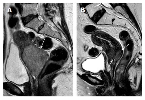
Figure 10 Advanced cervical cancer.
Sagittal T2-weighted images, before (A) and 2 mo after (B) radiation treatment. There is marked decrease in tumor size (> 50%), indicative of partial response to therapy. Residual tumor is seen as a high signal intensity area (arrows in B).
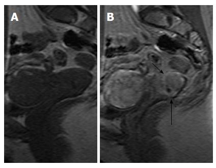
Figure 11 Dynamic contrast enhanced images, cervical cancer.
Sagittal pre- (A) and early arterial (30 s) post contrast (B) images. Cervical tumor borders are clearly delineated on the enhanced image (arrows in B). Note early arterial enhancement of cervical tumor in (B) synchronous with that of myometrium.
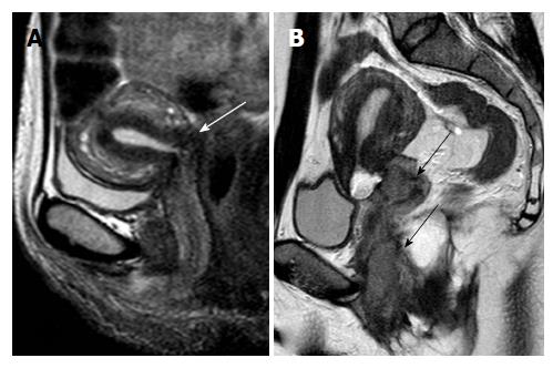
Figure 12 Post-operative sagittal T2-weighted images in two different patients treated with abdominal radical trachelectomy for early cervical cancer.
A: Normal postoperative appearance of the uterovaginal anastomosis (white arrow); B: Tumor relapse at the uterovaginal anastomosis extending to the lower vagina and pelvic floor (black arrows).
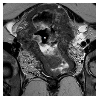
Figure 13 Bulky uterine cancer of indeterminate initial histology.
Oblique coronal T2-weighted image shows a large tumor involving the entire uterine corpus and cervix. Note retained secretions (black asterisk) and gas (white asterisk) in the endometrial cavity. When such extensive involvement is present, it may be difficult to determine the site of tumor origin (cervix or endometrium).
- Citation: Bourgioti C, Chatoupis K, Moulopoulos LA. Current imaging strategies for the evaluation of uterine cervical cancer. World J Radiol 2016; 8(4): 342-354
- URL: https://www.wjgnet.com/1949-8470/full/v8/i4/342.htm
- DOI: https://dx.doi.org/10.4329/wjr.v8.i4.342


