Copyright
©The Author(s) 2016.
World J Radiol. Feb 28, 2016; 8(2): 192-199
Published online Feb 28, 2016. doi: 10.4329/wjr.v8.i2.192
Published online Feb 28, 2016. doi: 10.4329/wjr.v8.i2.192
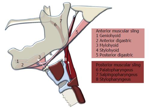
Figure 1 Two-sling mechanism for hyolaryngeal elevation in swallowing.
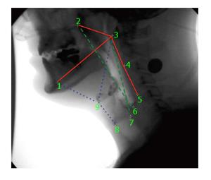
Figure 2 Pictured here are the nine coordinates mapping three skeletal levers (in red) and muscular slings displacing the hyolaryngeal complex with coordinates 1, 3, 8, 9 mapping the anterior muscular sling (in blue) and coordinates 2, 3, 6, 7 mapping the posterior muscular sling (in green).
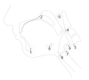
Figure 3 Procrustes fit of coordinates adjusts for differences in rotation and scale.
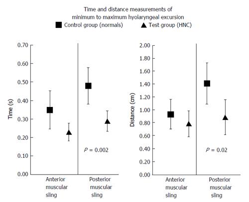
Figure 4 Significant differences between control and test groups.
HNC: Head and neck cancer.
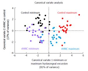
Figure 5 Canonical variate analysis showing differences in coordinate configuration plotted by canonical variate scores with classification variables highlighted.
rtHNC: Radiation therapy head and neck cancer.
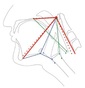
Figure 6 Eigenvectors indicating mechanics of hyolaryngeal elevation of the control group from minimum (dotted lines) to maximum (solid lines) excursion.
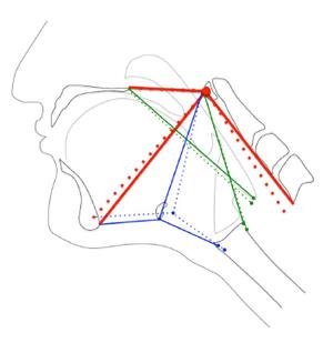
Figure 7 Eigenvectors indicating mechanics of hyolaryngeal elevation of the radiation therapy head and neck cancer group from minimum (dotted lines) to maximum (solid lines) excursion.
- Citation: Pearson Jr WG, Davidoff AA, Smith ZM, Adams DE, Langmore SE. Impaired swallowing mechanics of post radiation therapy head and neck cancer patients: A retrospective videofluoroscopic study. World J Radiol 2016; 8(2): 192-199
- URL: https://www.wjgnet.com/1949-8470/full/v8/i2/192.htm
- DOI: https://dx.doi.org/10.4329/wjr.v8.i2.192


