Copyright
©The Author(s) 2016.
World J Radiol. Feb 28, 2016; 8(2): 132-141
Published online Feb 28, 2016. doi: 10.4329/wjr.v8.i2.132
Published online Feb 28, 2016. doi: 10.4329/wjr.v8.i2.132
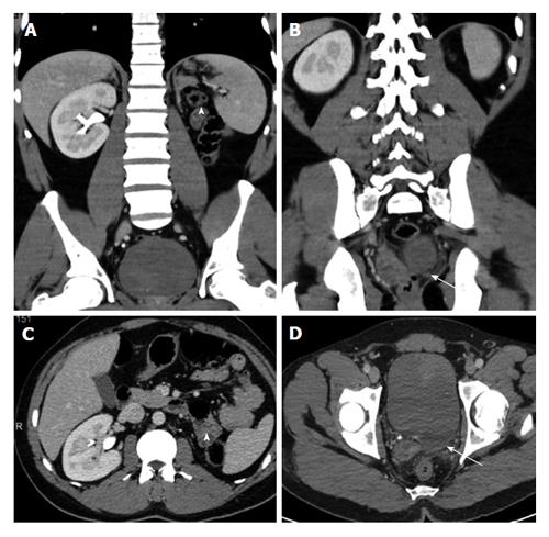
Figure 1 A 35-year-old man with left renal agenesis.
Coronal (A and B) and axial (C and D) contrast enhanced CT shows absent left kidney (arrowhead) and absent left seminal vesicle (arrow). CT: Computed tomography.
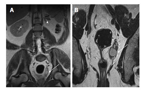
Figure 2 A 40-year-old man with left renal agenesis.
Coronal T2W magnetic resonance imaging abdomen (A) and pelvis (B) shows absent left kidney (arrowhead) and left seminal vesicle cyst (arrow).
Figure 3 Persistent fetal lobulations.
A: USG shows pseudomass from hypertrophied column of Bertin (arrow); B: USG shows Dromedary hump (arrow) from splenic impression (arrowhead); C: Coronal CT shows small LK with persistent fetal lobulations (arrow) inbetween the pyramids. CT: Computed tomography; USG: Ultrasonogram; LK: Left kidney.
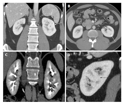
Figure 4 A 50-year-old woman with suspicious solid mass in left kidney on ultrasonogram.
Coronal (A and C), axial (B) and sagittal (D) computed tomography showing hypertrophied column of Bertin in interpolar region of LK (arrows) showing density and enhancement pattern similar to adjacent normal renal cortex. LK: Left kidney.
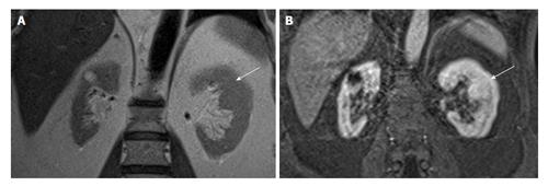
Figure 5 A 60-year-old man with suspicious mass in the left kidney on ultrasonogram.
Coronal T2W (A) and contrast enhanced T1W magnetic resonance imaging (B) shows hypertrophied column of Bertin in interpolar region of LK (arrows). LK: Left kidney.
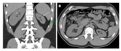
Figure 6 A 35-year-old man presented with right flank pain and incidental left renal hypoplasia.
Coronal (A) and axial (B) non-contrast computed tomography shows uniformly small left kidney with no scarring (arrow). Compensatory hypertrophy of right kidney seen with tiny calculi.
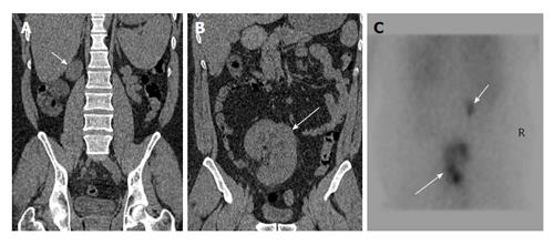
Figure 7 A 45-year-old man presenting with recurrent lower abdominal pain.
Coronal non-contrast computed tomography (A and B) shows hypoplastic right kidney (short arrow) and ectopic left kidney (long arrow). Nuclear scan (C) show small poorly functioning right kidney (short arrow) and ectopic left kidney (long arrow).
Figure 8 A 15-year-old girl with non-specific abdominal pain.
USG (A and B) shows normal right kidney and multiple non communicating cysts replacing left kidney. Nuclear scan (C) non functioning left kidney (arrows). USG: Ultrasonogram.
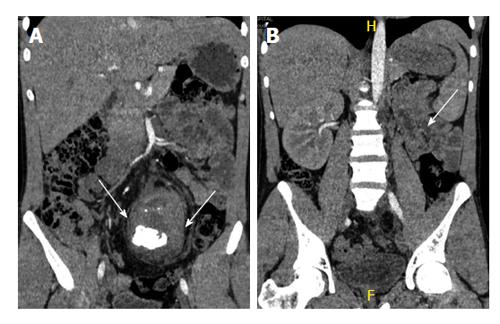
Figure 9 A 40-year-old man with long standing recurrent lower abdominal pain.
Coronal non-contrast computed tomography (A and B) shows ectopic left kidney in the pelvis with stag horn calculus causing hydronephrosis (arrows).
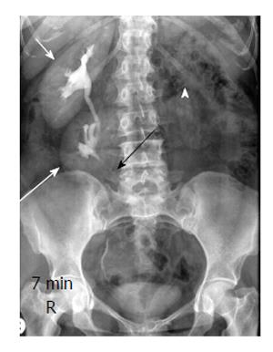
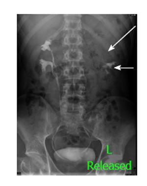
Figure 10 A 30-year-old man with crossed ectopia without fusion.
Intravenous urography shows crossed ectopia of left kidney (long arrow) below the normal right kidney (short arrow). Note ureter of ectopic left kidney crossing to left side (black arrow) and empty left renal fossa (arrowhead).
Figure 11 A 42-year-old man with crossed fused ectopia on the right side.
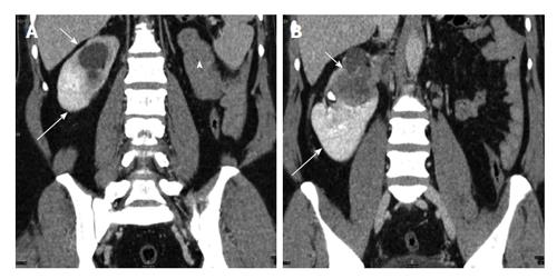
Coronal contrast enhanced computed tomography (A and B) shows empty left renal fossa (arrowhead) with crossed fused ectopic left kidney (long arrow) fused with the right kidney (short arrow) which shows infiltrating transitional cell carcinoma.
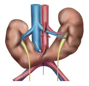
Figure 12 Schematic diagram showing the horse shoe kidneys due to the arrested migration of isthmus as the inferior mesenteric artery (white arrow) hooks over the isthmus (black arrow).
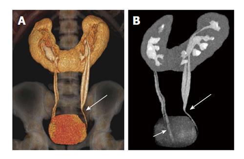
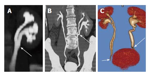
Figure 13 A 32-year-old woman with combined horse shoe kidney and duplex ureters.
Coronal volume rendered image (A) and Maximum intensity projection image (B) images show horse-shoe kidney with bilateral duplex ureters joining at the midureter level (long arrow) on left side and close to bladder (short arrow) on the right side.
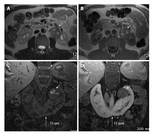
Figure 14 A 43-year-old man on follow up for horse shoe kidney.
Axial T2W (A) and T1W (B) MRI show horse shoe kidney (long arrow) with heterogenous mass in the left renal moiety showing T1 hyperintensity suggestive of blood products. Coronal T1 precontrast (C) and post contrast (D) MRI shows mild enhancement in the renal mass (short arrows). Histopathology came out as renal carcinoid. MRI: Magnetic resonance imaging.
Figure 15 A 25-year-old man with bilateral duplex kidneys and “drooping lily” sign on the left side.
One hour delayed radiograph after computed tomography urogram shows bilateral duplex system with hydronephrotic left upper moiety (long arrow) displacing the lower moiety (short arrow).
Figure 16 Different forms of duplex kidneys based on the level of fusion.
A: Bifid renal pelvis; B: Y shaped ureter; C: V shaped ureter (short arrow). Also seen left lower ureteric stricture (long arrow).
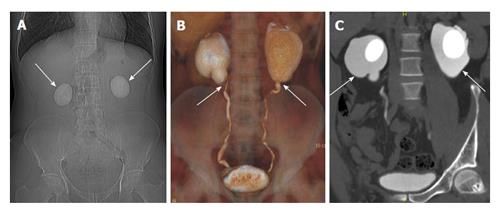
Figure 17 A 24-year-old man with bilateral flank pain.
A: Computed tomography scannogram shows bilateral large round renal calculi; B: Coronal Volume rendered image shows narrowing at bilateral Pelvi-ureteric junction; C: Coronal maximum intensity projection image shows bilateral ballooning of pelvis (arrows) with large round renal pelvic calculi.
- Citation: Ramanathan S, Kumar D, Khanna M, Al Heidous M, Sheikh A, Virmani V, Palaniappan Y. Multi-modality imaging review of congenital abnormalities of kidney and upper urinary tract. World J Radiol 2016; 8(2): 132-141
- URL: https://www.wjgnet.com/1949-8470/full/v8/i2/132.htm
- DOI: https://dx.doi.org/10.4329/wjr.v8.i2.132


