Copyright
©The Author(s) 2016.
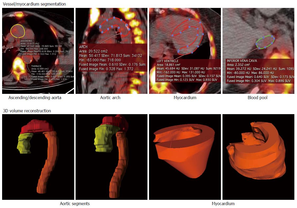
Figure 1 Volume of interest placement and 3-dimensional rendering of the vascular segments and of the myocardium.
Volumes of interest were constructed by placing sequential region of interest on the transaxial CT slices, carefully adjusting the edges so as to include the entire vessel/myocardium (top panels). The figure depicts the 3D rendering of the three aortic segments and of the heart (bottom panels). 3D: 3-dimensional; CT: Computed tomography.
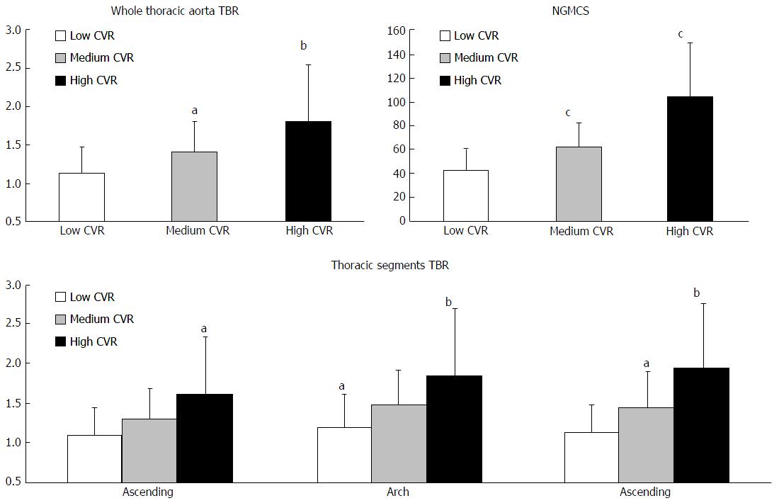
Figure 2 Target-to-background ratio differences in cardiovascular risk classes.
The figure highlights the average TBR values for the whole thoracic aorta (top left), the myocardium (top right) and the three different thoracic aorta segments (bottom). aP < 0.05; bP < 0.01; cP < 0.001. TBR: Target-to-background ratio; CVR: Cardiovascular risk; NGMCS: Normalized global molecular calcification score.
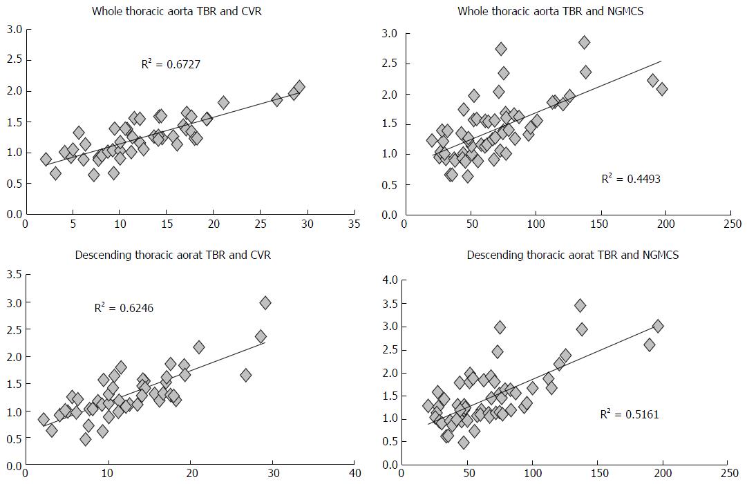
Figure 3 Correlation between fluoride uptake and clinical cardiovascular risk.
A strong correlation was noted between thoracic aorta NaF uptake and CVR, but also between aortic and myocardial uptake (top panels). Among the thoracic aorta subsegments, descending aorta provided the strongest correlation (bottom panels). TBR: Target-to-background ratio; CVR: Cardiovascular risk; NGMCS: Normalized global molecular calcification score.
- Citation: Fiz F, Morbelli S, Bauckneht M, Piccardo A, Ferrarazzo G, Nieri A, Artom N, Cabria M, Marini C, Canepa M, Sambuceti G. Correlation between thoracic aorta 18F-natrium fluoride uptake and cardiovascular risk. World J Radiol 2016; 8(1): 82-89
- URL: https://www.wjgnet.com/1949-8470/full/v8/i1/82.htm
- DOI: https://dx.doi.org/10.4329/wjr.v8.i1.82


