©The Author(s) 2015.
World J Radiol. Jul 28, 2015; 7(7): 180-183
Published online Jul 28, 2015. doi: 10.4329/wjr.v7.i7.180
Published online Jul 28, 2015. doi: 10.4329/wjr.v7.i7.180
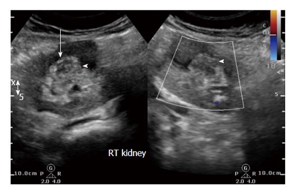
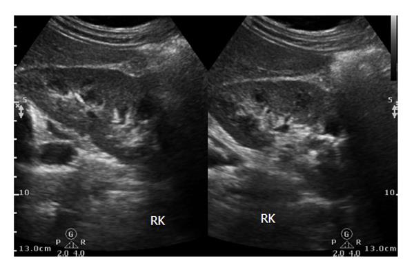
Figure 1 Initial ultrasound image reveals heteroechoic indeterminate renal sinus/renal pelvic lesion (white arrow).
Doppler revealed negligible vascularity.
Figure 2 Composite of non contrast axial MDCT-post contrast corticomedullary and post contrast nephrographic phases reveals poorly enhancing isodense lesion within the renal pelvis exhibiting calcific specks (white arrow) and hypo-attenuating lesion along its lateral aspect (red arrows).
Subtle thickening of the urothelial walls is present.
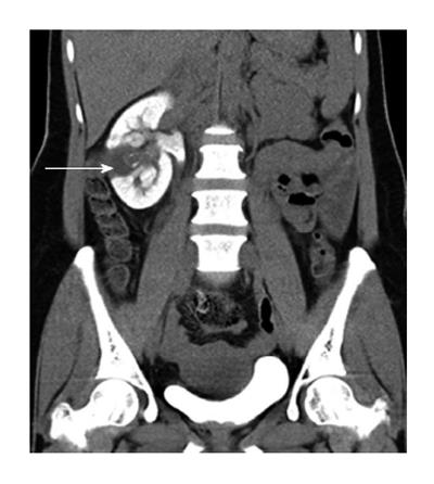
Figure 3 Delayed oblique coronal multiplanar reformat MDCT image shows the lesion as a filling defect within the collecting system.
The lesion and laterally placed cortical cyst are inseparable. Additionally, persistent nephrogram is noted.
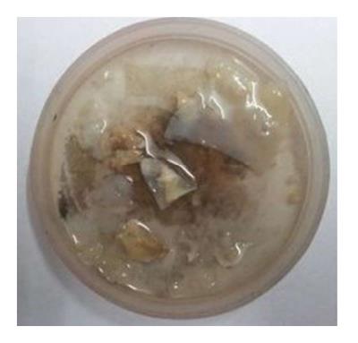
Figure 4 Photograph of the fleshy membranes passed spontaneously by the patient few days after ureteroscopy.
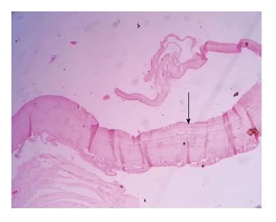
Figure 5 Microscopy (magnification 100 ×) revealed characteristic hydatid lamellated layer (arrow) with occasional brood capsules.
Figure 6 Follow up ultrasound revealed normal right kidney.
- Citation: Bhaya A, Shinde AP. Isolated renal hydatid presenting as a complex renal lesion followed by spontaneous hydatiduria. World J Radiol 2015; 7(7): 180-183
- URL: https://www.wjgnet.com/1949-8470/full/v7/i7/180.htm
- DOI: https://dx.doi.org/10.4329/wjr.v7.i7.180


