Copyright
©The Author(s) 2015.
World J Radiol. Oct 28, 2015; 7(10): 336-342
Published online Oct 28, 2015. doi: 10.4329/wjr.v7.i10.336
Published online Oct 28, 2015. doi: 10.4329/wjr.v7.i10.336
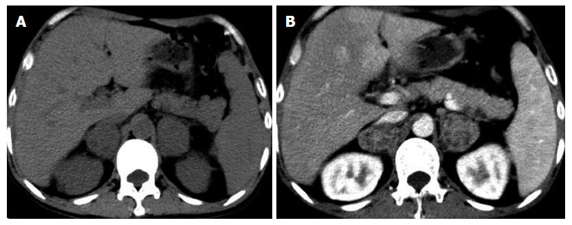
Figure 1 A 53-year-old man who has fatigue, pigmentation of skin and loss weight in last five months with primary adrenal insufficiency due to adrenal tuberculosis.
The unenhanced (A) and contrast-enhanced (B) CT scans reveal the mass-like enlargement of the bilateral adrenals with multifocal peripheral enhancement. CT: Computed tomography.
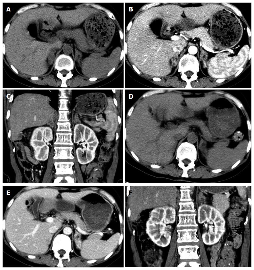
Figure 2 A 52-year-old woman who has anorexia, daytime somnolence and sweating in last three months with primary adrenal insufficiency due to adrenal tuberculosis.
The unenhanced (A) and contrast-enhanced (B) CT scans reveal the mass-like enlargement of bilateral adrenals, but its contours are preserved with multifocal peripheral enhancement on axial (B) and coronary (C) reformed images. Three months after antituberculous therapy, the axial (D and E) and coronary (F) images demonstrate the initially enlarged adrenal glands become small with homogeneous density in the center, and decrease of probability of the presence of peripheral rim enhancement. CT: Computed tomography.
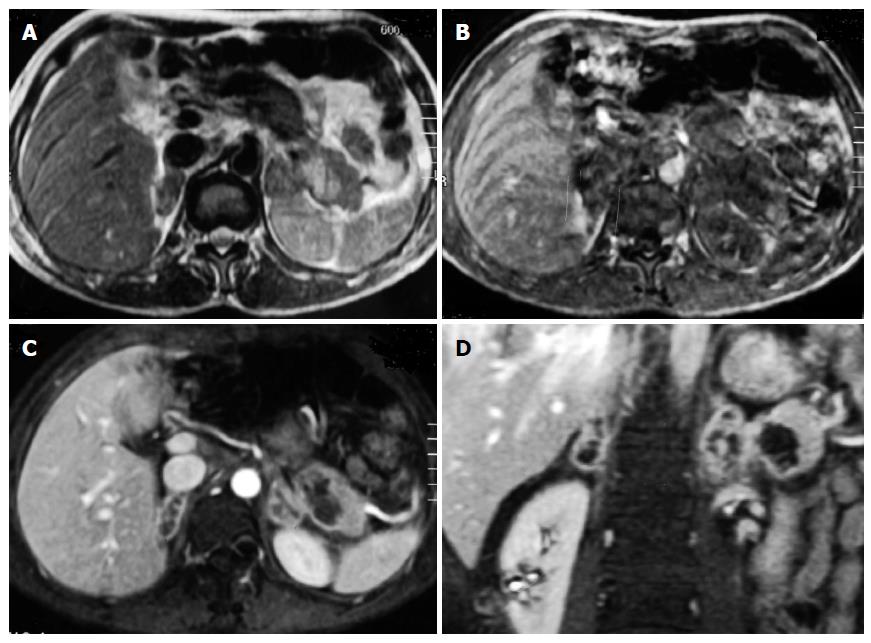
Figure 3 A 44-year-old man who has hypoglycemia and low blood pressure with primary adrenal insufficiency due to adrenal tuberculosis.
MRI scans reveal the mass-like enlargement of bilateral adrenal glands on axial T1- weighted image (A) and T2-weighted image (B), but its contours are preserved with peripheral enhancement on contrast-enhanced axial (C) and coronal (D) T1-weighted image. MRI: Magnetic resonance imaging.
Figure 4 A 51-year-old man with pigmentation of skin and blood electrolyte abnormalities due to primary adrenal insufficiency due to adrenal tuberculosis.
The unenhanced (A) and contrast-enhanced (B) CT scans illustrate the enlargement of the left adrenal gland with peripheral enhancement. This patient underwent adrenal pathology biopsy, and photomicrograph (C) shows central caseous necrosis surrounded by granulomatous inflammatory cells (× 40). CT: Computed tomography.
- Citation: Huang YC, Tang YL, Zhang XM, Zeng NL, Li R, Chen TW. Evaluation of primary adrenal insufficiency secondary to tuberculous adrenalitis with computed tomography and magnetic resonance imaging: Current status. World J Radiol 2015; 7(10): 336-342
- URL: https://www.wjgnet.com/1949-8470/full/v7/i10/336.htm
- DOI: https://dx.doi.org/10.4329/wjr.v7.i10.336


