Copyright
©The Author(s) 2015.
World J Radiol. Oct 28, 2015; 7(10): 306-318
Published online Oct 28, 2015. doi: 10.4329/wjr.v7.i10.306
Published online Oct 28, 2015. doi: 10.4329/wjr.v7.i10.306
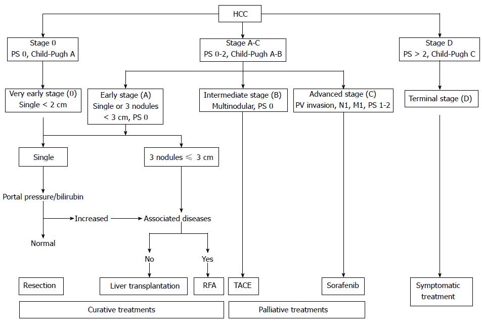
Figure 1 Flow chart of Barcelona Clinic Liver Cancer based management guidelines for hepatocellular carcinoma[2].
HCC: Hepatocellular carcinoma; RFA: Radiofrequency ablation; TACE: Transarterial chemoembolization.
Figure 2 Axial computed tomography images before and after radiofrequency ablation shows an arterial enhancing lesion (arrow, A).
That is replaced by a hypodense area without any enhancement following radiofrequency ablation (arrow, B).
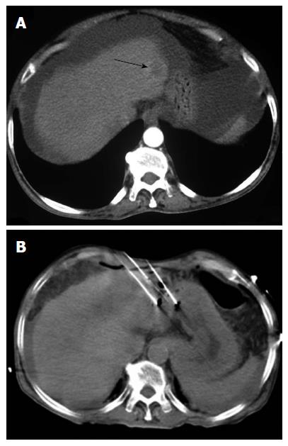
Figure 3 Irreversible electroporation for an early stage hepatocellular carcinoma in left lobe (arrow) (A and B).
Figure 4 Computed tomography 1 mo after irreversible electroporation in the same patient as Figure 4.
No residual enhancement is seen (arrow).
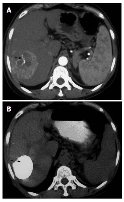
Figure 5 Trans-arterial chemoembolisation of a large hepatocellular carcinoma in right lobe of liver (arrow, A).
Following transarterial chemoembolization, there is uniform distribution of lipiodol the lesion (arrow, B).
- Citation: Kalra N, Gupta P, Chawla Y, Khandelwal N. Locoregional treatment for hepatocellular carcinoma: The best is yet to come. World J Radiol 2015; 7(10): 306-318
- URL: https://www.wjgnet.com/1949-8470/full/v7/i10/306.htm
- DOI: https://dx.doi.org/10.4329/wjr.v7.i10.306


