©2014 Baishideng Publishing Group Inc.
World J Radiol. Jun 28, 2014; 6(6): 374-380
Published online Jun 28, 2014. doi: 10.4329/wjr.v6.i6.374
Published online Jun 28, 2014. doi: 10.4329/wjr.v6.i6.374
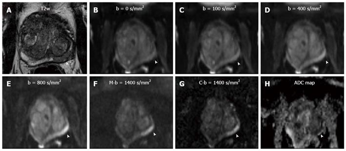
Figure 1 Axial T2-weighted image of an 88 years old patient with a PSA level of 8.
2 ng/mL and negative findings upon digital rectal examination. T2w shows a focal hypointense lesion in the posterolateral left midgland pz (arrowhead in A). Measured b-values of 0, 100, 400 and 800 s/mm² (arrowheads in B, C, D and E, respectively) do not depict the suspected lesion with sufficient contrast to background prostate. M-b1400 shows good lesion-to-prostate contrast (arrowhead in F). C-b1400 depicts the lesion with the same lesion-to-prostate contrast and a slightly improved demarcation (arrowhead in G). The ADC value of the suspicious lesion was as low as 648 × 10-3 mm/s² [ADC map (H)].
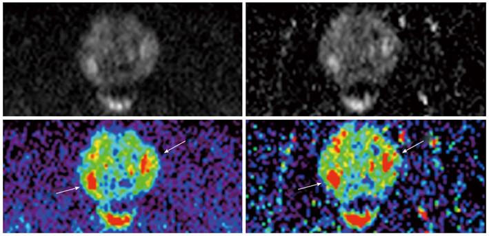
Figure 2 M-b1400 (left) and C-b1400 (right) images of a 63 years old patient with elevated PSA levels, at corresponding axial slice position.
Region-of-interests were placed in the prostate and in the bladder content to obtain signal intensity-ratios to estimate signal-to-noise-ratio. Calculated b-value images show higher SI-ratios and more anatomical image details (i.e., bladder contour).
Figure 3 M-b1400 (left column) and C-b1400 (right column) images of the same patient.
Demarcation of two oval hyperintense suspicious lesions is very good on both images (arrows).
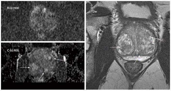
Figure 4 Images of a 68 years old patient with elevated PSA levels and signs of benign prostate hyperplasia.
M-b1400 (upper image on the left) was acquired with reduced image quality due to distortion artifacts. The C-b1400 (below) in corresponding slice position and identical window level shows an improved image quality with demarcation of several lesions that were rated as benign benign prostate hyperplasia-nodules taking into account T2w (right image) and dynamic contrast-enhanced imaging.
-
Citation: Bittencourt LK, Attenberger UI, Lima D, Strecker R, Oliveira A, Schoenberg SO, Gasparetto EL, Hausmann D. Feasibility study of computed
vs measured high b-value (1400 s/mm²) diffusion-weighted MR images of the prostate. World J Radiol 2014; 6(6): 374-380 - URL: https://www.wjgnet.com/1949-8470/full/v6/i6/374.htm
- DOI: https://dx.doi.org/10.4329/wjr.v6.i6.374


