©2014 Baishideng Publishing Group Inc.
World J Radiol. Jun 28, 2014; 6(6): 223-229
Published online Jun 28, 2014. doi: 10.4329/wjr.v6.i6.223
Published online Jun 28, 2014. doi: 10.4329/wjr.v6.i6.223
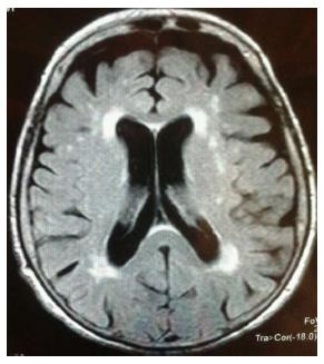
Figure 1 White matter hyperintensities as assessed by magnetic resonance images.
White matter hyperintensities (WMHs) appear as hyperintense signals on T2-weighted magnetic resonance images and represent ependymal loss and differing degrees of myelination. These lesions can be related to a wide variety of clinical conditions and pathophysiological processes. WMHs, depending on the localization, are commonly classified as periventricular hyperintensities (PWMHs) or deep white matter hyperintensities (DWMHs). DWMHs were identified as having mainly a vascular aetiology whereas PWMHs could be due to ependymal loss, differing degrees of myelination and cerebral ischemia. WMHs are reported to be commonly associated with older age, and cardiovascular risk factors such as hypertension and diabetes.
- Citation: Serafini G, Gonda X, Rihmer Z, Girardi P, Amore M. White matter abnormalities: Insights into the pathophysiology of major affective disorders. World J Radiol 2014; 6(6): 223-229
- URL: https://www.wjgnet.com/1949-8470/full/v6/i6/223.htm
- DOI: https://dx.doi.org/10.4329/wjr.v6.i6.223


