©2014 Baishideng Publishing Group Inc.
World J Radiol. May 28, 2014; 6(5): 203-209
Published online May 28, 2014. doi: 10.4329/wjr.v6.i5.203
Published online May 28, 2014. doi: 10.4329/wjr.v6.i5.203
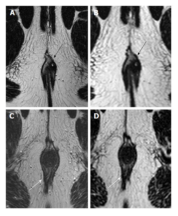
Figure 1 Two different patients imaged with both 2D (A and C) and 3D T2 turbo spin-echo (B and D) images.
In most patients, such as a 30-year-old male with intersphincteric fistula (black arrows), depictions were clear on both 2D (A) and 3D (B). However, in the 39-year-old male depicted in images C and D, the fistula (white arrows) entering dorsally was noticed on 2D images (C), but not those that were 3D (D). Note that there is a seton partially visible inside the tract on the 2D image.
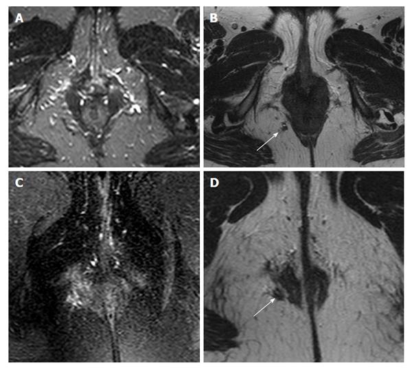
Figure 2 A 45-year-old female imaged with short tau inversion recovery (A), 2D T2 turbo spin-echo (B and D), and T2 with fat saturation (C).
The small vessels mask the extrasphincteric fistula (white arrows) that was corrected after being noticed on T2 turbo spin-echo. T2 with fat saturation (C) was unable to detect the fistula or correctly identify its relationship to the sphincter complex (D).
Figure 3 A 18-year-old male with complex fistula with two tracts (black arrows).
Demonstration of these tracts for the surgeon on both oblique axial (A) and sagittal 2D TSE images (B) is perhaps more difficult than a reformatted 3D T2 TSE image (C). TSE: Turbo spin-echo.
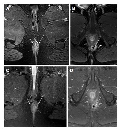
Figure 4 Two male subjects, one 39-year old (A and B) and the other 52 years old (C and D), imaged with 3D T1 with fat saturation (A and C) and T1 turbo spin-echo with fat saturation (B and D).
The 39-year-old male (A and B) has an intersphincteric fistula treated with a seton (long white arrows). Both the fistula and seton inside the fistula and luminal part (short arrows) are seen more clearly on 3D images. The 52-year-old has a small intersphincteric abscess cavity (white arrows C and D), which is shown well on both sequences. However the contrast between fat and muscle is poorer on turbo spin-echo compared to 3D T1, making it much easier to correctly appreciate the relationship between fistula/abscess and the surrounding sphincter.
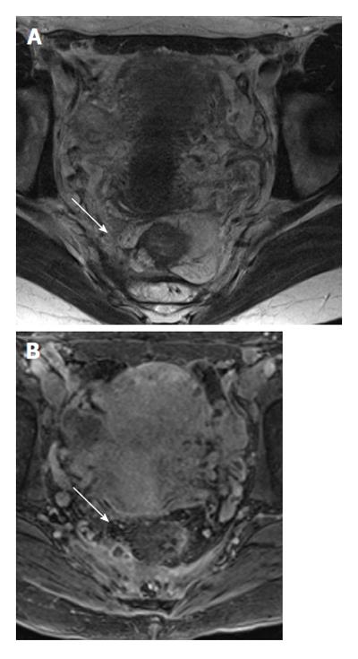
Figure 5 A 20-year-old with intersphincteric fistula (white arrows) treated with seton image.
A: 2D T2 turbo spin-echo; B: 3D T1 weighted prepared gradient echo sequence with fat saturation. The seton is seen clearly on THRIVE, as well as the inflammation, but not on T2. However, the intersphincteric course of the fistula is clearer on T2, while the extent of inflammation obscures the thin external sphincter.
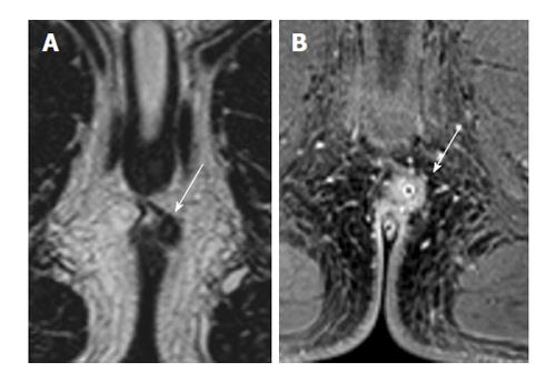
Figure 6 A 45-year-old female with fistula extending above the levator ani muscles (supralevator) and forming an abscess (white arrows).
The signal intensity of the fat and fluid are the same on T2 without fat saturation, and the abscess can easily be missed on T2 weighted images (A). However, the marked inflammation on 3D T1 post-contrast clearly demonstrates the abscess (B).
- Citation: Torkzad MR, Ahlström H, Karlbom U. Comparison of different magnetic resonance imaging sequences for assessment of fistula-in-ano. World J Radiol 2014; 6(5): 203-209
- URL: https://www.wjgnet.com/1949-8470/full/v6/i5/203.htm
- DOI: https://dx.doi.org/10.4329/wjr.v6.i5.203


