Copyright
©2014 Baishideng Publishing Group Co.
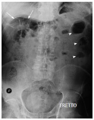
Figure 1 Upright abdominal plain film.
Some air-fluid levels can be appreciated in the left flank (arrow-heads) with air-driven distension of small bowel loops (arrows) in the right hypochondrium and normal fecal content in the right colon.
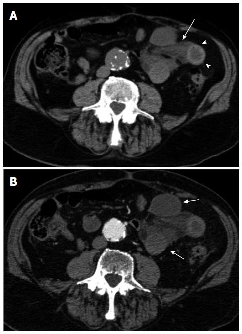
Figure 2 Multi-detector computed tomography.
Unenhanced (A) and contrast-enhanced (B) axial images of the abdomen are shown. In A, a cluster of fluid-filled dilated small bowel loops can be appreciated in the left flank along with the evidence of mesenteric fluid (arrow). A mural hematoma can also be appreciated (arrow-heads). In B lack of enhancement of the intestinal wall of the small bowel loops is depicted as a result of bowel wall ischemia.
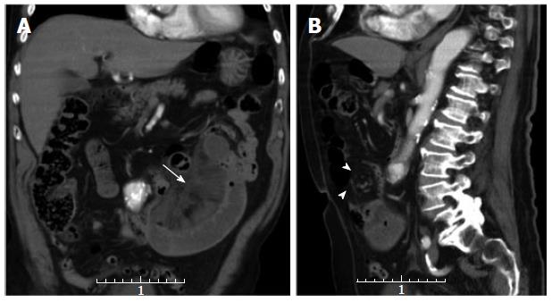
Figure 3 Multi-detector contrast-enhanced computed tomography.
Coronal (A) and sagittal (B) reformatted 5 mm thick images are shown. In (A) a cluster of ischemic jejunal loops is depicted in the left flank along with mesenteric fluid (arrow). In (B) swirling of the mesenteric vessels (arrow-heads) is depicted. The computed tomography finding (whirl sign) was prospectively considered consistent with a small bowel volvulus which was not confirmed at surgery.
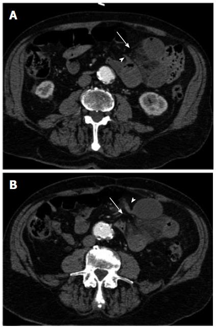
Figure 4 Multi-detector contrast-enhanced computed tomography.
At retrospective analysis, a beak-like appearance (arrow-heads) of the closely apposed afferent (A) and efferent (B) jejunal loops can be appreciated along with the hernial orifice (arrow) in the periphery of the omentum. The peripheral location of the herniated loops and the absence of an overlying omental fat can also be appreciated.
- Citation: Camera L, Gennaro AD, Longobardi M, Masone S, Calabrese E, Vecchio WD, Persico G, Salvatore M. A spontaneous strangulated transomental hernia: Prospective and retrospective multi-detector computed tomography findings. World J Radiol 2014; 6(2): 26-30
- URL: https://www.wjgnet.com/1949-8470/full/v6/i2/26.htm
- DOI: https://dx.doi.org/10.4329/wjr.v6.i2.26


