©2013 Baishideng Publishing Group Co.
World J Radiol. Jun 28, 2013; 5(6): 241-247
Published online Jun 28, 2013. doi: 10.4329/wjr.v5.i6.241
Published online Jun 28, 2013. doi: 10.4329/wjr.v5.i6.241
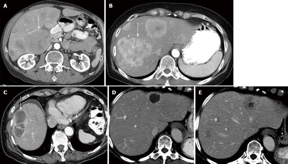
Figure 1 Sixty-one-year-old female with metastatic islet cell carcinoma.
A: Transverse LAVA images of the abdomen following Gd contrast administration. The lesions in the liver (arrow) are hyperenhancing lesions relative to the liver; B: Computed tomography image of the abdomen following IV contrast. The lesions in the liver are hypoenhancing relative to the liver (arrow).
Figure 2 Computed tomography image of the abdomen following IV contrast.
A: 56-year-old male with islet cell cancer. The lesion in the liver is iso enhancing relative to the liver (arrow); B: 58-year-old male with islet cell cancer. The lesions in the liver are less than 50% necrosis and hyper; C: 77-year-old male with islet cell cancer. The lesion in the liver is more than 50% necrosis; D: 58-year-old male with islet cell cancer. The lesion in the liver is more than 50% necrosis in the post-treatment enhancing relative to the liver; E: 58-year-old male with islet cell cancer. The lesion in the liver demonstrates less than 50% necrosis on the pre-treatment images.
- Citation: Neperud J, Mahvash A, Garg N, Murthy R, Szklaruk J. Can imaging patterns of neuroendocrine hepatic metastases predict response yttruim-90 radioembolotherapy? World J Radiol 2013; 5(6): 241-247
- URL: https://www.wjgnet.com/1949-8470/full/v5/i6/241.htm
- DOI: https://dx.doi.org/10.4329/wjr.v5.i6.241


