©2013 Baishideng Publishing Group Co.
World J Radiol. Jun 28, 2013; 5(6): 229-240
Published online Jun 28, 2013. doi: 10.4329/wjr.v5.i6.229
Published online Jun 28, 2013. doi: 10.4329/wjr.v5.i6.229
Figure 1 Dysplastic nodules in a cirrhotic liver.
Early enhancement with persistent vascularity of 2 nodules (solid arrows) in a patient with chronic liver disease. The synchronous B mode image shows the same hypoechoic ill defined nodules (dotted arrows).
Figure 2 Hemangioma detected incidentally.
A: Contrast ultrasound showing central filling of a rounded non echogenic lesion in the liver on B mode ultrasound (arrow); B: Synchronous B mode image of the lesion, not appearing like a classical hemangioma (arrow); C: T1 weighted contrast enhanced axial section of the liver on magnetic resonance imaging with peripheral enhancing rim of the lesion (arrow); D: Subsequent centripetal filling up of the lesion (arrow) on equilibrium phase of contrast enhanced T1 weighted axial sections of the liver.
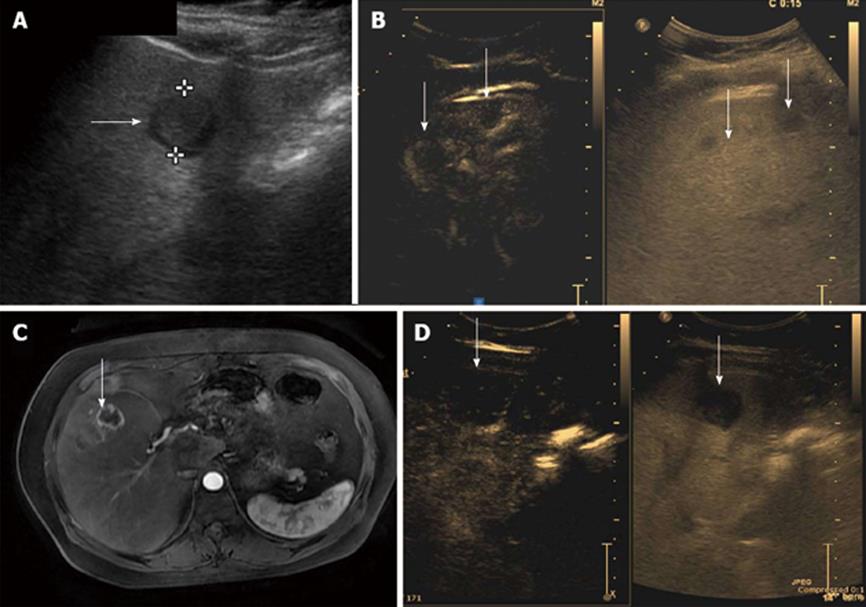
Figure 3 Metastases from neuroendocrine tumor.
A: Hypoechoic target like lesion on B mode ultrasound on routine ultrasound (arrow) B: Contrast enhanced ultrasound (CEUS) shows at least 2 lesions (arrows) with early arterial peripheral hypervascularity with synchronous B mode image of the lesions; C: T1 weighted contrast enhanced axial section of the liver on magnetic resonance imaging with hypervascular peripheral enhancing rim of the lesion in arterial phase (arrow); D: CEUS shows washout on delayed scans (arrow).
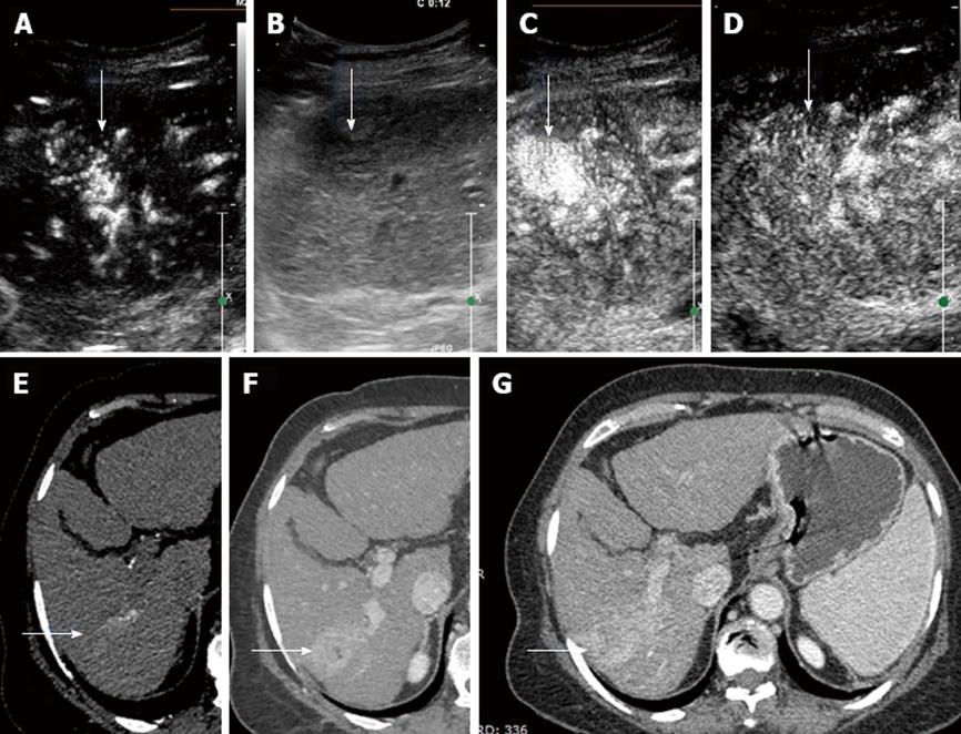
Figure 4 Hepatocellular carcinoma with classical enhancement pattern on contrast enhanced ultrasound in a 50-year-old patient with hepatitis B infection, detected to have a mass on routine screening.
A: Early arterial enhancement of the mass on contrast enhanced ultrasound (CEUS) (arrow); B: B mode synchronous image of the mass lesion (arrow); C: Rapid contrast enhancement of the lesion on CEUS (arrow); D: Early washout of contrast within the lesion (arrow) on CEUS; E: Arterial phase enhancement of the lesion on triple phase computer tomography (CT) scan (arrow); F: Portal venous phase on dynamic CT showing lesion enhancement (arrow); G: Equilibrium Phase on CT showing lesion contrast washout (arrow).
Figure 5 Hepatocellular carcinoma with enhancing tumor thrombus on contrast enhanced ultrasound.
A: Hypoechoic mass in the liver on routine B mode ultrasound (arrow); B: On injection of ultrasound contrast early arterial enhancement of the mass (arrow); C: Peripheral capsule formation of the mass with increased enhancement (arrow); D: Washout of the contrast on delayed scanning (arrow); F: Persistent portal vein enhancing thrombus on contrast scan with (arrow); E: Echogenic thrombus seen within portal vein lumen; F: Dynamic computer tomography scan of the same patient showing hypervascular mass with tumor thrombus (arrow).
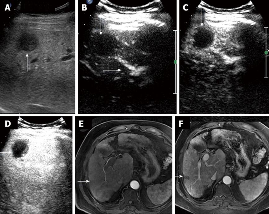
Figure 6 Post radiofrequency ablation lesion assessed with contrast ultrasound followed by dynamic magnetic resonance imaging showing no significant residual tumor.
A: Hypoechoic lesion in segment VII of liver on B mode ultrasound (arrow); B: Post contrast ultrasound minimal peripheral enhancement (solid arrow) with feeding vessel sign (dotted arrow); C: Contrast ultrasound image showing mild peripheral enhancement without obvious residual tumor (arrow); D: Delayed scan post contrast on contrast enhanced ultrasound shows lesion as a hypoechoic defect (arrow); E: Arterial phase on axial T1 weighted magnetic resonance imaging (MRI) of liver without lesion hypervascularity (arrow); F: Portal venous phase on axial T1 weighted scans of liver on MRI show segment VII hypointense non enhancing lesion (arrow).
Figure 7 Bar chart showing the various lesions assessed and comparison of the diagnostic specificity of contrast enhanced ultrasound and reference standards (computer tomography/magnetic resonance imaging).
CT/MRI: Computer tomography/magnetic resonance imaging; CEUS: Contrast enhanced ultrasound; HCC: Hepatocellular carcinoma; CLD: Chronic liver disease; RFA: Radio frequency ablation; TACE: Trans-arterial chemo embolization.
- Citation: Laroia ST, Bawa SS, Jain D, Mukund A, Sarin S. Contrast ultrasound in hepatocellular carcinoma at a tertiary liver center: First Indian experience. World J Radiol 2013; 5(6): 229-240
- URL: https://www.wjgnet.com/1949-8470/full/v5/i6/229.htm
- DOI: https://dx.doi.org/10.4329/wjr.v5.i6.229


