Copyright
©2013 Baishideng Publishing Group Co.
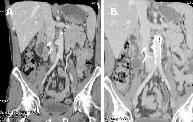
Figure 1 An endovascular repair of a saccular aneurysm of the infra-renal aorta.
A: Original Saccular aneurysm. Etiology was never defined; B: Post endovascular repair.
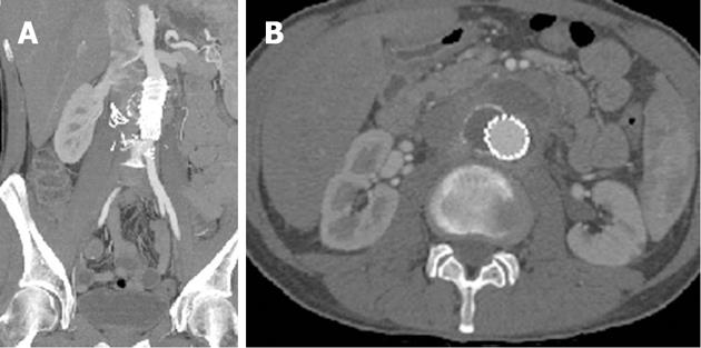
Figure 2 Fluid and phlegmon surrounding the graft.
A: Coronal view when patient presented demonstrating residual sac and new left sided fluid; B: Axial view demonstrating peri-graft fluid and phlegmon.
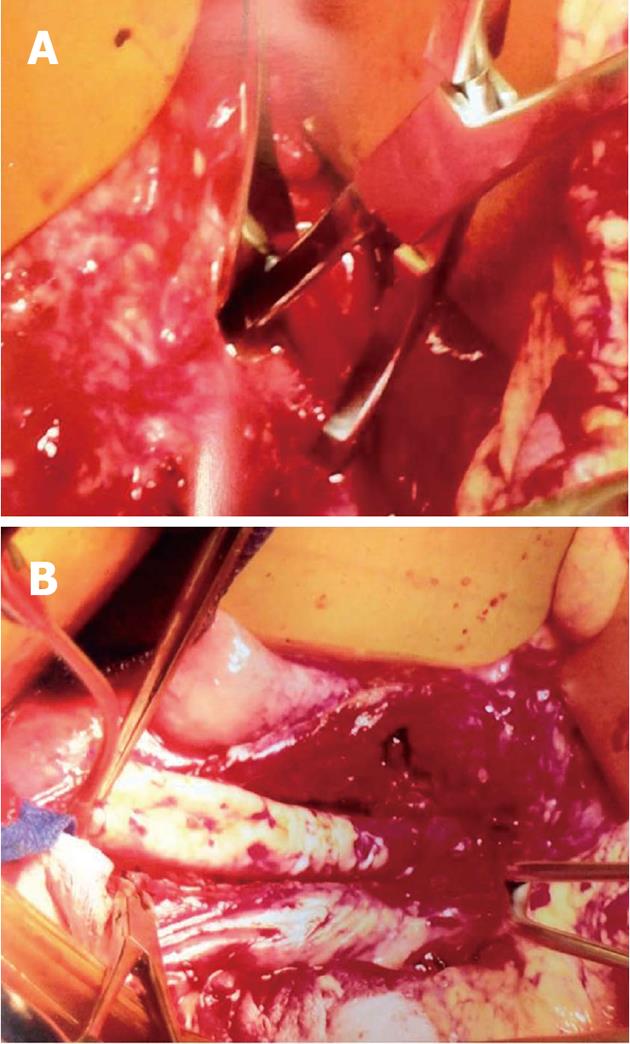
Figure 3 Operative pictures.
A: Operative picture demonstrating the dense phlegmon with proximal clamp below renal arteries; B: Operative picture with proximal anastomosis complete and after debridement of the phlegmon and aortic wall.
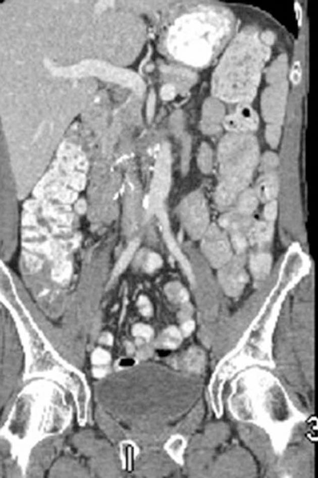
Figure 4 Computer tomographic angiography at one year follow up.
-
Citation: Teso D, Williams S, Karmy-Jones R.
Pasturella multicoda infection of an abdominal aortic endograft. World J Radiol 2013; 5(1): 17-19 - URL: https://www.wjgnet.com/1949-8470/full/v5/i1/17.htm
- DOI: https://dx.doi.org/10.4329/wjr.v5.i1.17


