©2012 Baishideng Publishing Group Co.
World J Radiol. Sep 28, 2012; 4(9): 391-400
Published online Sep 28, 2012. doi: 10.4329/wjr.v4.i9.391
Published online Sep 28, 2012. doi: 10.4329/wjr.v4.i9.391
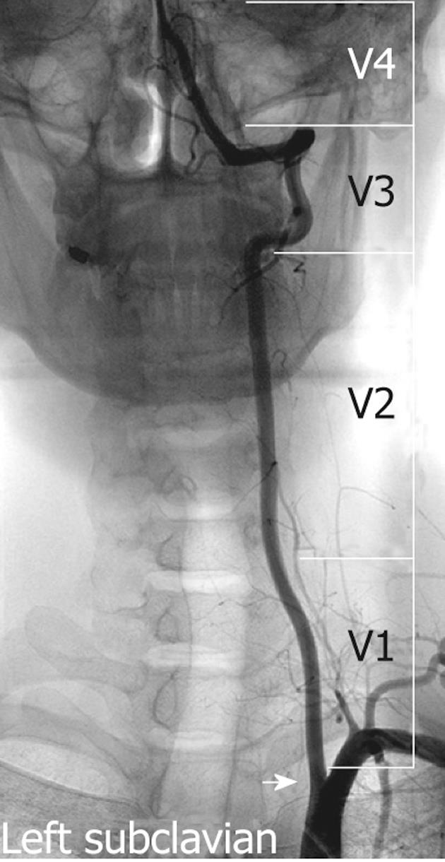
Figure 1 Non-subtracted left subclavian angiography demonstrates the vertebral artery orifice (arrow), the three extracranial segments (V1-3) and one intracranial segment (V4).
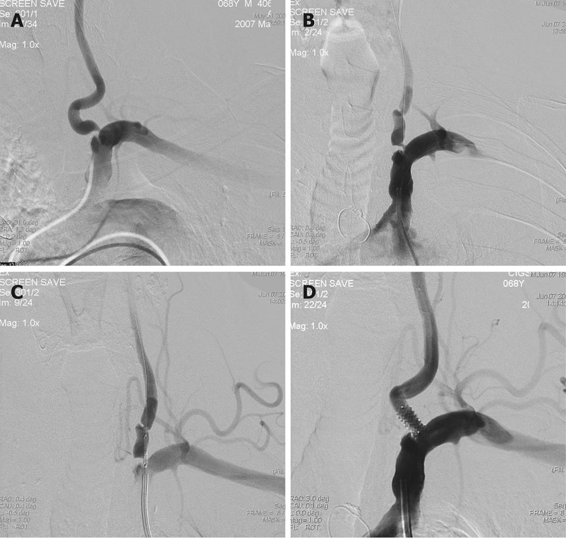
Figure 2 Classical approach for vertebral artery stenting.
A-C: Left subclavian angiography posterior-anterior (PA) view shows ulcerated short segment significant stenosis in the origin of the left vertebral artery (A), placement of the microguidewire to the left vertebral artery and microguidewire to the subclavian artery as a guidewire (B) and correct placement of the coronary balloon-expandable stent in the stenosed segment (C); D: Post-stent angiography shows good opposition of the stent and well opening of the stenosed segment.
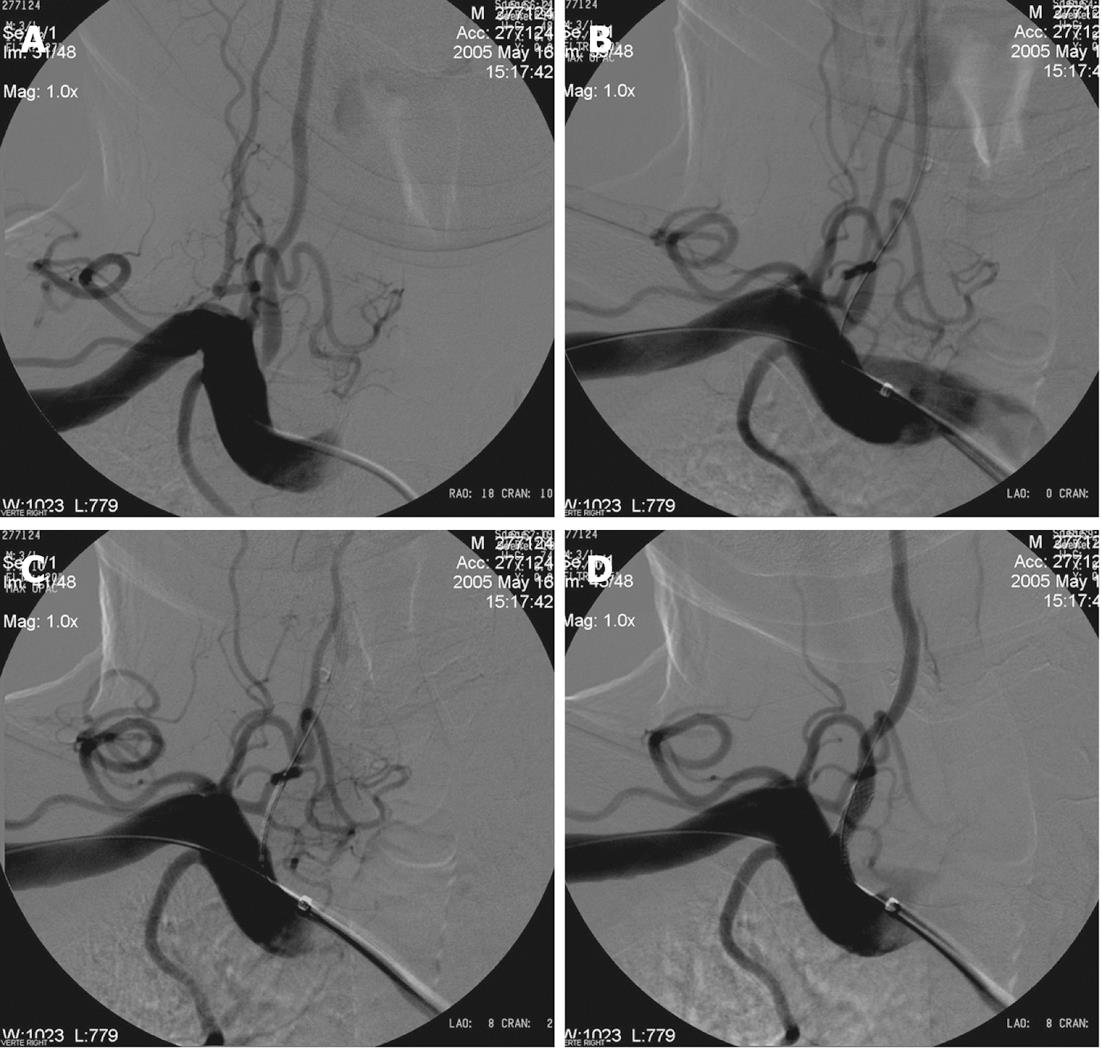
Figure 3 Buddy wire technique for tortuous subclavian arteries.
A: Right subclavian angiography posterior-anterior (PA) view shows significant concentric stenosis of the right VA origin; B: Right subclavian angiography shows placement of the stent to the right vertebral artery (VA) and one microguidewire to the subclavian artery as a buddy wire; C: Right subclavian angiography after placement of the stent shows temporary occlusion of the right VA and correct placement of the stent; D: Right subclavian angiography after opening of the stent shows good anatomical result.
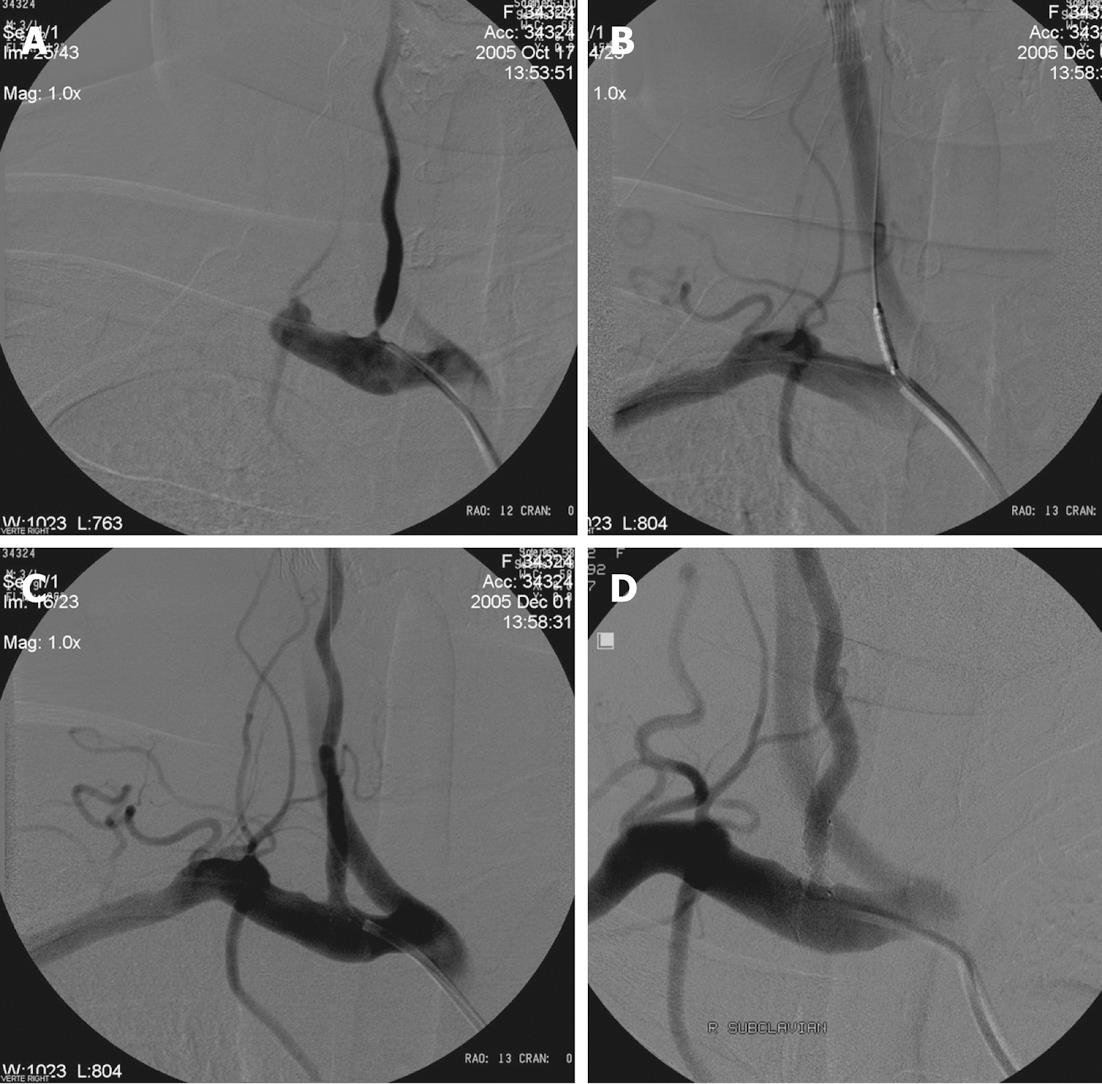
Figure 4 Radiological follow-up.
A: Right subclavian angiography posterior-anterior (PA) view shows significant stenosis of the right vertebral artery origin; B: Placement of the coronary balloon-expandable stent in the stenosed segment; C: After opening of the stent angiography shows well opposition of the stent and lack of residual stenosis; D: Eighteen months control angiography shows patency of the right VA origin and stent.
- Citation: Kocak B, Korkmazer B, Islak C, Kocer N, Kizilkilic O. Endovascular treatment of extracranial vertebral artery stenosis. World J Radiol 2012; 4(9): 391-400
- URL: https://www.wjgnet.com/1949-8470/full/v4/i9/391.htm
- DOI: https://dx.doi.org/10.4329/wjr.v4.i9.391


