©2011 Baishideng Publishing Group Co.
World J Radiol. Dec 28, 2011; 3(12): 279-288
Published online Dec 28, 2011. doi: 10.4329/wjr.v3.i12.279
Published online Dec 28, 2011. doi: 10.4329/wjr.v3.i12.279
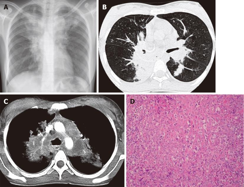
Figure 1 Hodgkin’s lymphoma.
A 23-year-old woman with a four-mo history of dry cough and chest pain. A: Chest X-ray shows mediastinal widening and upper lobe parenchymal opacities; B and C: Contrast-enhanced computed tomography confirms lymphadenopathy involving the mediastinum and infiltrative masses in the bilateral upper lobes; D: Photomicrograph (HE stain). Nodular sclerosing HL lymph node. Fibrous bands divide the lymphoid infiltrate into nodules and contain Hodgkin cells.
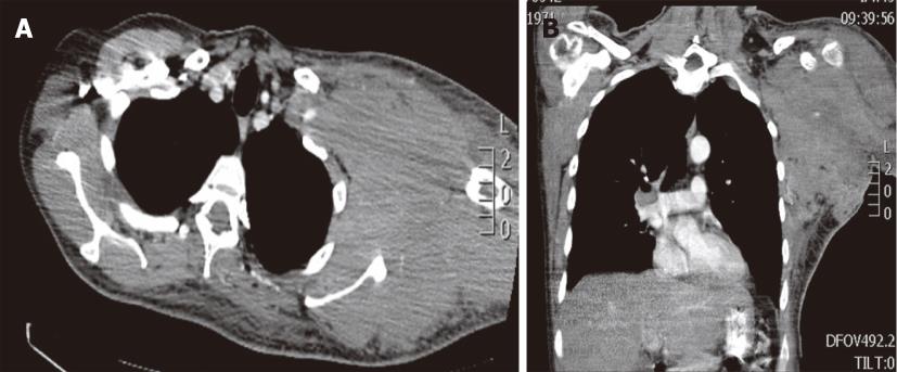
Figure 2 Burkitt lymphoma in a 38-year-old male with a left axilla mass.
Contrast-enhanced computed tomography, axial (A) and coronal (B) images demonstrates a bulky soft tissue mass involving the axilla, subpectoral and subscapular spaces.
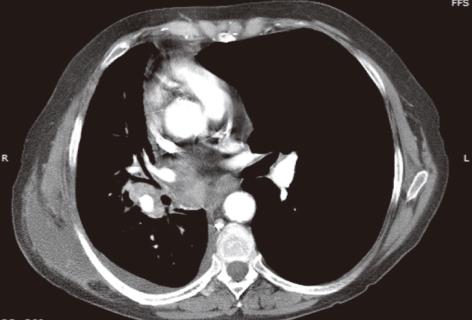
Figure 3 Post-transplant proliferative disease in a 50-year-old male post-right lung transplant.
Contrast-enhanced computed tomography shows an infiltrative right hilar mass surrounding the right lower lobe bronchus and involving the subcarinal region. Ipsilateral small pleural effusion is also noted.
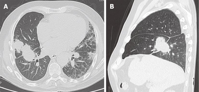
Figure 4 Post-right lung transplant post-transplant proliferative disease in a 68-year-old male with pulmonary fibrosis.
Non-contrast computed tomography, axial (A) and sagittal images (B) demonstrates an irregular mass in the right lung involving the middle lobe and lower lobe extending across the major fissure.
Figure 5 Lymphomatoid granulomatosis in a young adult male with acquired immunodeficiency syndrome.
Initial chest computed tomography (CT) (A) demonstrates a round solid mass in the right lower lobe. After surgical resection follow-up CT (B) 4 mo later shows recurrent disease with new multiple nodules and cavitation in the right lower lobe.
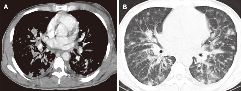
Figure 6 Kaposi sarcoma in a 37-year-old male with acquired immunodeficiency syndrome.
Contrast enhanced computed tomography of the chest, mediastinal window (A) and lung window (B) images demonstrate innumerable peribronchovascular and peripheral pulmonary nodules throughout the bilateral lungs. Enhancing skin lesions and bilateral pleural effusion are also noted.
Figure 7 Multicentric Castleman’s disease in a 40-year-old male with acquired immunodeficiency syndrome.
Contrast-enhanced chest computed tomography, axial images at two different levels (A, B) reveal numerous enhancing abnormally enlarged lymph nodes in the axillae and mediastinum.
Figure 8 Recurrent respiratory papillomatosis in a 27-year-old patient.
Contrast-enhanced computed tomography, axial (A, B) and coronal (C) images. Numerous nodules and cystic lesion are seen in the bilateral lungs as well as a large lobulated mass partially obstructing the distal trachea (arrows).
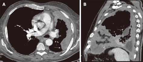
Figure 9 Malignant mesothelioma in a 60-year-old male with progressive chest pain.
Contrast-enhanced computed tomography, axial (A) and sagittal (B) images show an irregular and slightly lobulated soft tissue density mass extensively involving the pleural surface of the left lung including the major fissure (arrows).
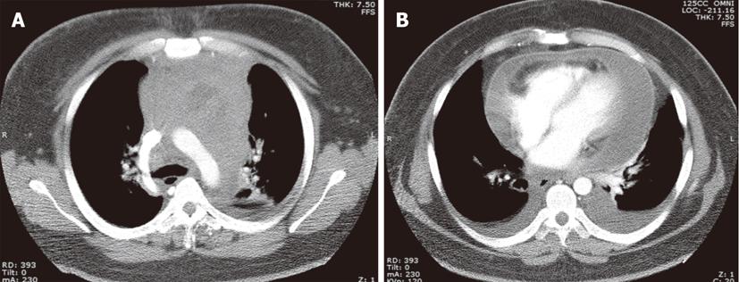
Figure 10 Adult T cell leukemia in a 24-year-old male.
Contrast-enhanced computed tomography at the level of the aortic arch (A) and mid-ventricular level (B) demonstrates a large mediastinal mass with extensive pericardial involvement and bilateral pleural effusions.
Figure 11 Non-Hodgkin lymphoma in two different patients with acquired immunodeficiency syndrome.
A: Contrast-enhanced computed tomography (CT) at the level of the AP window demonstrates a large mediastinal mass with areas of cavitation with air-fluid levels (arrows); B: Contrast-enhanced CT reveals a large mediastinal mass with low-density foci consistent with tumoral necrosis. There is small left sided pleural effusion in both cases.
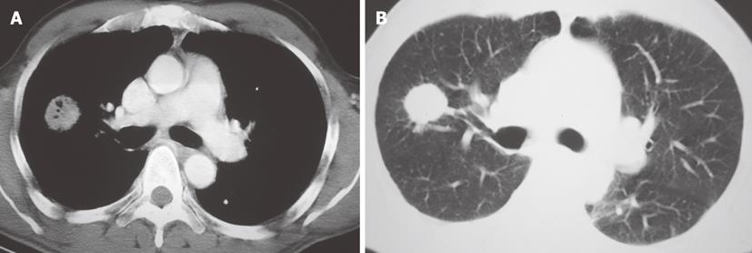
Figure 12 Contrast enhanced chest CT.
A: Mediastinum window, axial image at the level of the pulmonary arteries demonstrates a soft tissue density mass with air-bronchogram in the right lung. B: Lung window image at the same level shows the spiculated contour of the lung mass.
- Citation: Restrepo CS, Chen MM, Martinez-Jimenez S, Carrillo J, Restrepo C. Chest neoplasms with infectious etiologies. World J Radiol 2011; 3(12): 279-288
- URL: https://www.wjgnet.com/1949-8470/full/v3/i12/279.htm
- DOI: https://dx.doi.org/10.4329/wjr.v3.i12.279


