©2010 Baishideng Publishing Group Co.
World J Radiol. Sep 28, 2010; 2(9): 354-357
Published online Sep 28, 2010. doi: 10.4329/wjr.v2.i9.354
Published online Sep 28, 2010. doi: 10.4329/wjr.v2.i9.354
Figure 1 Non-enhanced cranial computed tomography of a 58-year-old female patient with a hematocrit of 58%.
Note that all depicted intracranial arteries and veins appear hyperdense (curved arrows). No contrast agent was applied prior to the scan.
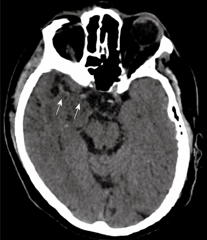
Figure 2 Pseudohyperdense right middle cerebral artery (arrows) due to underlying infection of the temporal lobe.
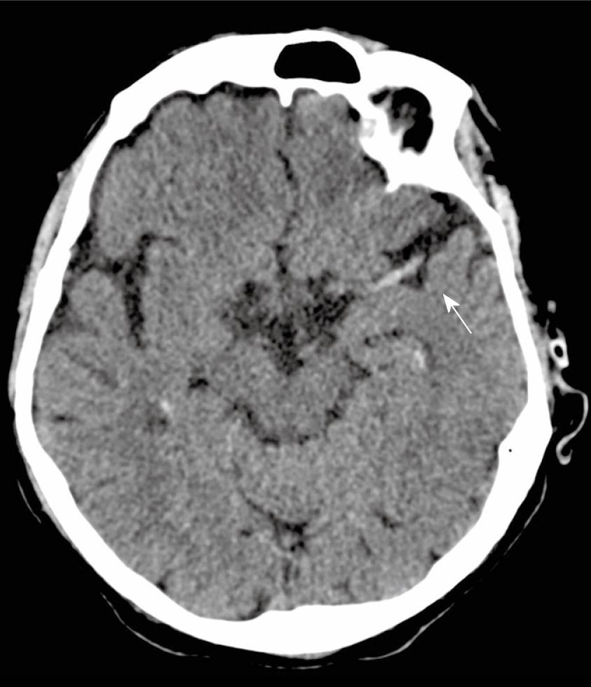
Figure 3 Hyperdense left middle cerebral artery sign (arrow) in a patient presenting with signs of left hemispheric stroke.
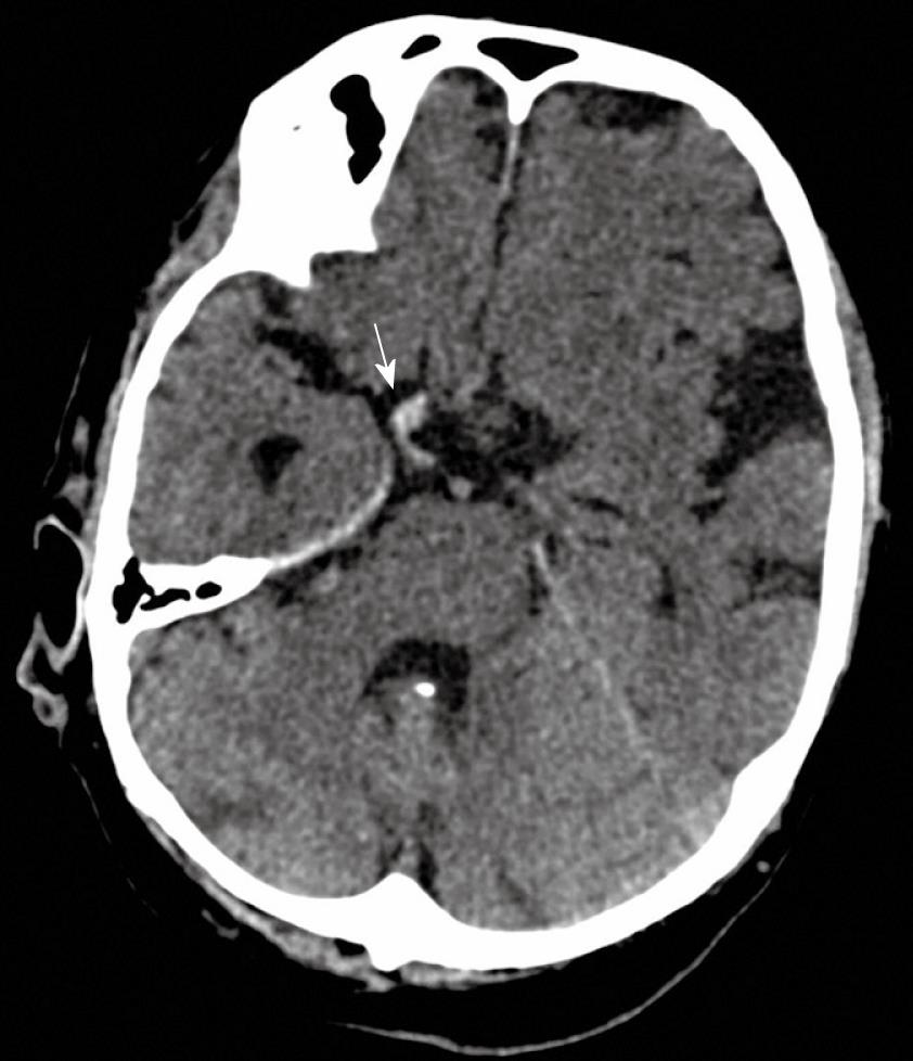
Figure 4 Hyperdense left anterior cerebral artery (arrow) in a patient who presented with right-sided hemiparesis.
Figure 5 Hyperdense right fetal posterior cerebral artery (arrow) in a patient who presented with left sided homonymous hemianopia and left sided hemiparesis.
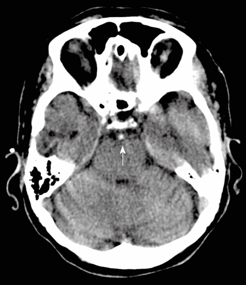
Figure 6 Hyperdense basilar artery (arrow) in a patient who was found unconscious.
- Citation: Jensen-Kondering U, Riedel C, Jansen O. Hyperdense artery sign on computed tomography in acute ischemic stroke. World J Radiol 2010; 2(9): 354-357
- URL: https://www.wjgnet.com/1949-8470/full/v2/i9/354.htm
- DOI: https://dx.doi.org/10.4329/wjr.v2.i9.354


