Copyright
©2010 Baishideng Publishing Group Co.
World J Radiol. May 28, 2010; 2(5): 159-165
Published online May 28, 2010. doi: 10.4329/wjr.v2.i5.159
Published online May 28, 2010. doi: 10.4329/wjr.v2.i5.159
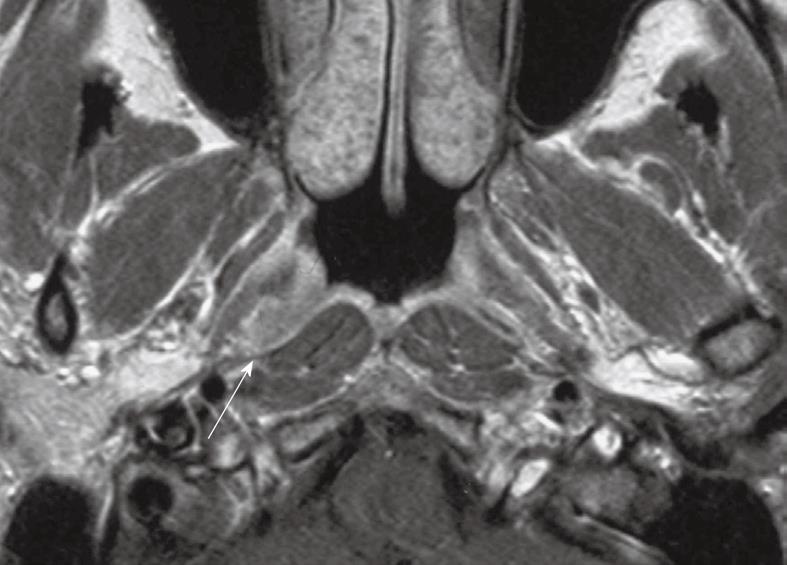
Figure 1 Axial post-contrast T1 weighted magnetic resonance imaging (MRI) showing a small nasopharyngeal carcinoma within the right lateral pharyngeal recess (arrow).
This is a frequent site for early cancer.
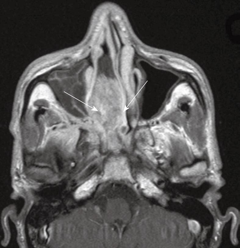
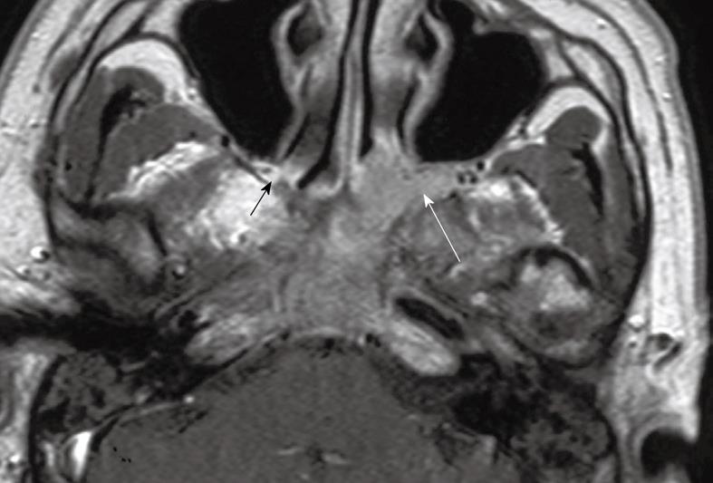
Figure 2 Axial post-contrast T1 weighted MRI showing a bulky nasopharyngeal carcinoma with gross extension into the right nasal cavity (arrows).
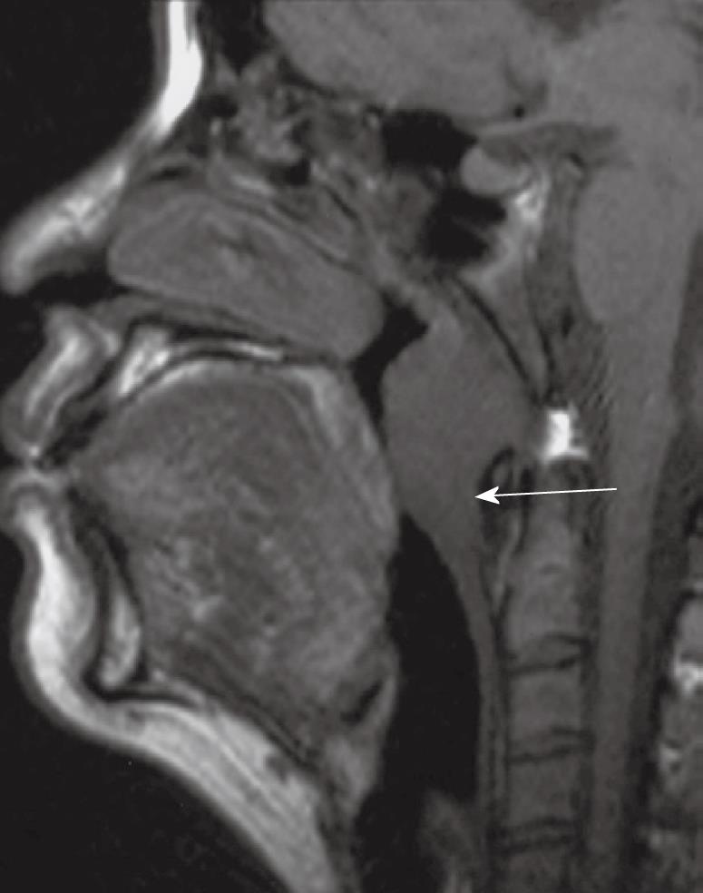
Figure 3 Sagittal T1 weighted MRI showing a bulky nasopharyngeal carcinoma with inferior extension, crossing the C1/2 level, along the posterior wall into the oropharynx (arrow).
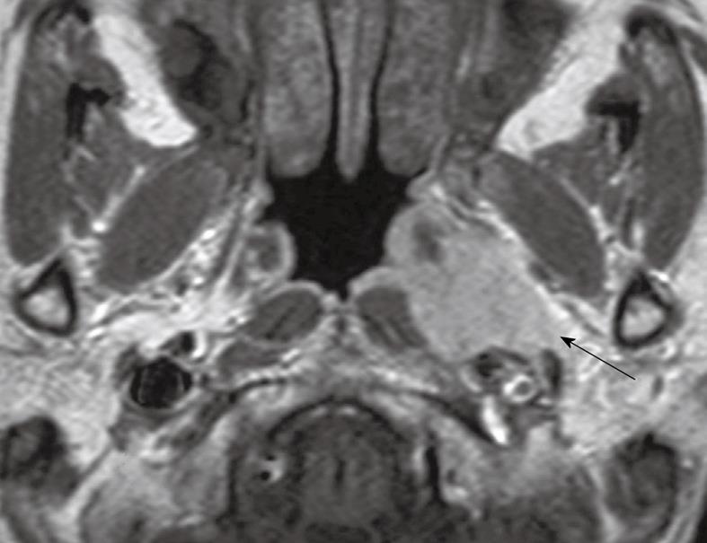
Figure 4 Axial post-contrast T1 weighted MRI showing a nasopharyngeal carcinoma directly infiltrating into the left parapharyngeal space (arrow).
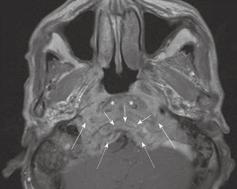
Figure 5 Axial post-contrast T1-weighted MRI showing a nasopharyngeal carcinoma with gross posterior retropharyngeal extension in the longus capitis muscles (asterisks), prevertebral space (long arrows), clivus (short arrows) and posterior cranial fossa (broken arrows).
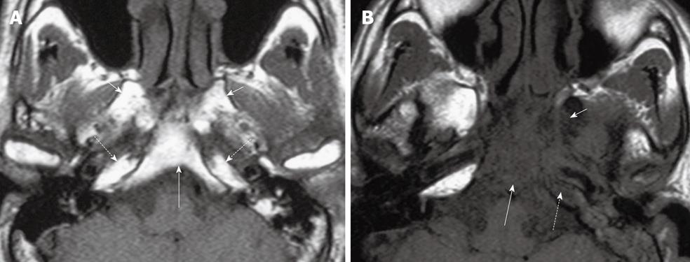
Figure 6 Axial T1 weighted MRI of the skull base showing five key bony sites to check for tumor invasion.
A: Normal skull base showing T1W weighted signal of normal fatty bone marrow within the clivus (long arrow), bilateral pterygoid bases (short arrows) and petrous apices (broken arrows); B: Abnormal skull base showing loss of normal high T1 weighted signal due to tumor invasion of the clivus (long arrow), left pterygoid base (short arrow) and left petrous apex (broken arrow).
Figure 7 Coronal post-contrast T1 weighted MRIs of nasopharyngeal carcinoma in three patients illustrating tumor extension in the foramina from the anterior (A) to the posterior (C) skull base.
A: The right foramen rotundum and vidian canal (solid long and short arrows, respectively); B: Left foramen ovale (solid arrow); C: Left foramen lacerum (solid arrow). The uninvolved foramina on the contralateral sides are indicated by broken arrows.
Figure 8 Axial post-contrast T1 weighted MRI showing contiguous extension of nasopharyngeal carcinoma into the left pterygopalatine fossa which is expanded (long arrow).
Compare this with the normal hyperintense fat signal in the narrow pterygopalatine fossa on the contralateral side (short arrow).
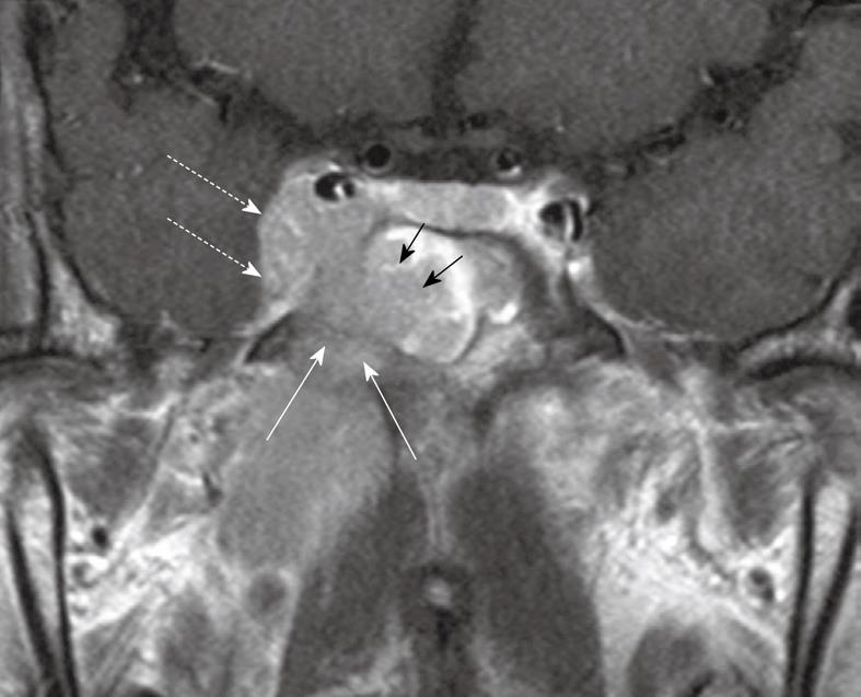
Figure 9 Coronal post-contrast T1 weighted MRI showing a nasopharyngeal carcinoma with direct infiltration through the sphenoid body (long arrows) into the sphenoid sinus (short arrows) and right cavernous sinus (broken arrows).
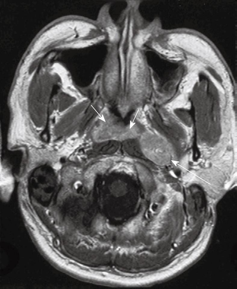
Figure 10 Axial post-contrast T1 weighted MRI showing a nasopharyngeal carcinoma (short arrows) and a grossly enlarged metastatic left retropharyngeal lymph node at the level of the nasopharynx (long arrow).
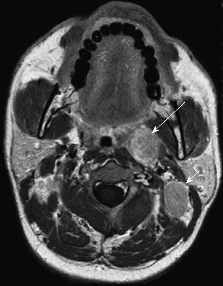
Figure 11 Axial post-contrast T1 weighted MRI in a patient with nasopharyngeal carcinoma showing an enlarged left sided metastatic retropharyngeal node deep to the oropharynx (long arrow).
An enlarged metastatic left upper internal jugular node posterior to the left internal jugular vein (level IIB) is also present which is a very common site of nodal metastases (short arrow).
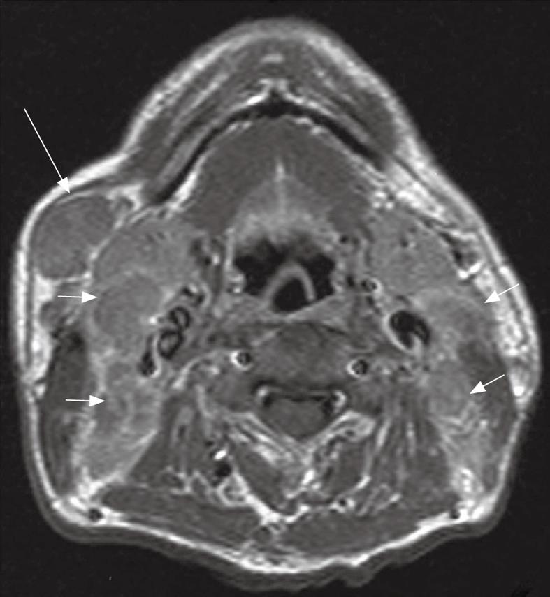
Figure 12 Axial post-contrast T1 weighted MRI in a patient with nasopharyngeal carcinoma showing bulky metastatic nodes in the internal jugular chains (short arrows) and right submandibular region (long arrow).
- Citation: King AD, Bhatia KSS. Magnetic resonance imaging staging of nasopharyngeal carcinoma in the head and neck. World J Radiol 2010; 2(5): 159-165
- URL: https://www.wjgnet.com/1949-8470/full/v2/i5/159.htm
- DOI: https://dx.doi.org/10.4329/wjr.v2.i5.159


