
















Copyright: ©Author(s) 2026.
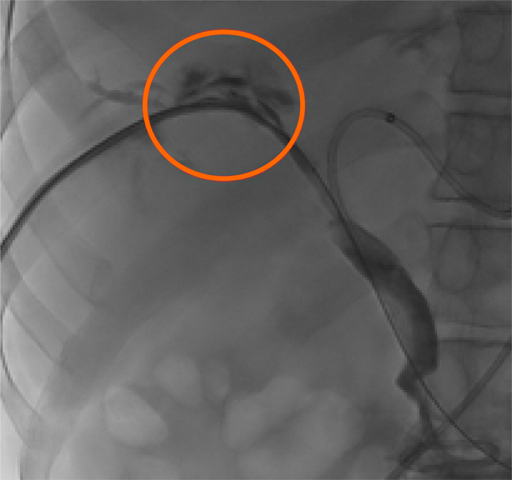
Figure 1 A 55-year-old male with malignant biliary obstruction (cholangiocarcinoma Klatskin 4).
The “pull-back cholangiogram” depicts an arterio-biliary fistula (orange circle).
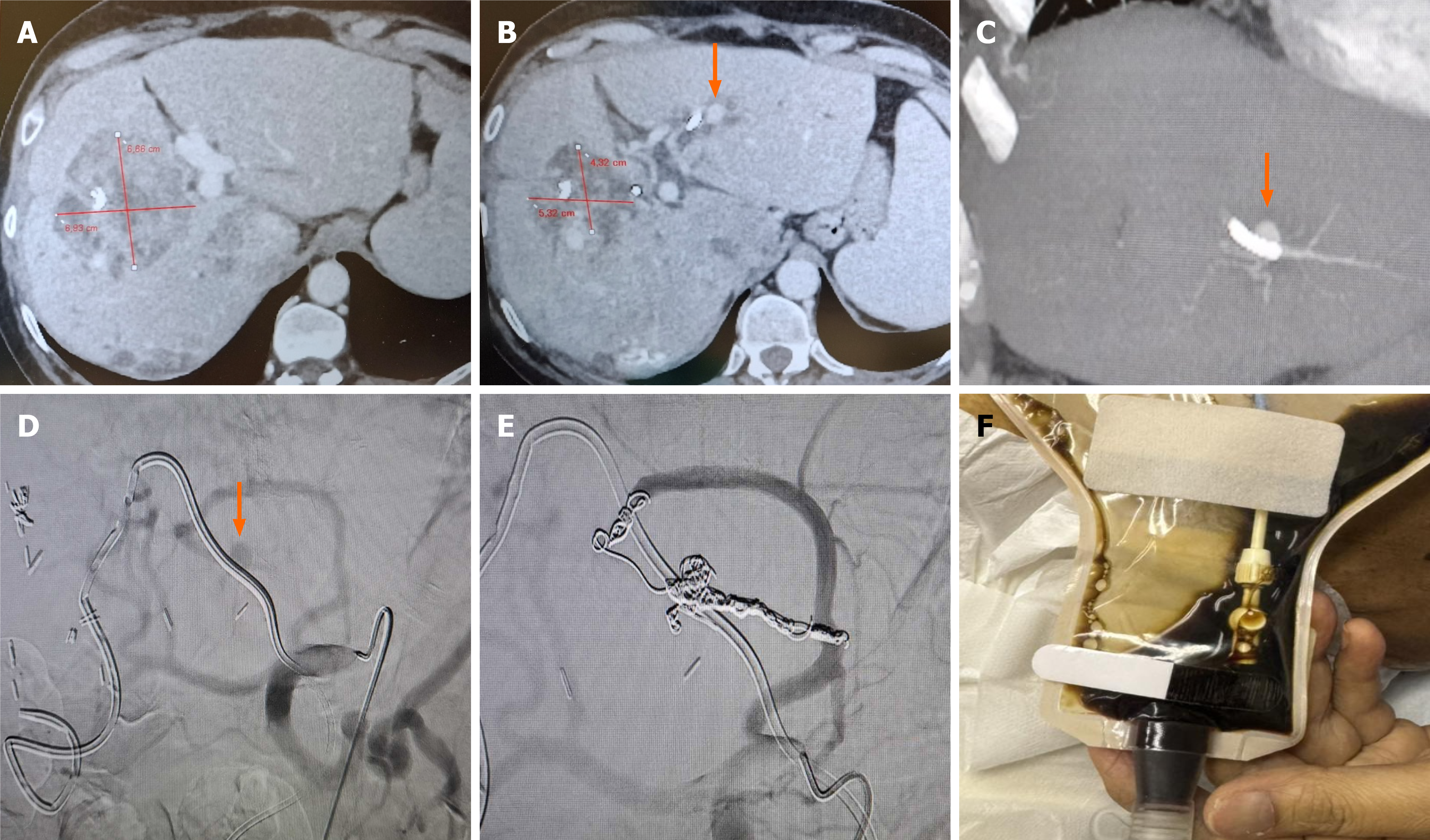
Figure 2 A 59-year-old female patient with obstructive jaundice due to three colorectal liver metastasis (max diameter of largest lesion 7 cm) underwent biliary drainage as to enable radioembolization therapy.
A: Access was obtained from the left lobe due to extensive right lobe metastatic disease. Selective Internal Radiation Therapy was successfully performed. However, 10 weeks after Selective Internal Radiation Therapy the patient reported blood within the percutaneous transhepatic biliary drainage collective bag. Following urgent admission hemobilia was diagnosed (right quadrant pain and melena); B and C: Computed tomography angiography depicted a pseudoaneurysm in proximity to the percutaneous biliary catheter (arrows), as well as good response to the radioembolization (treated lesions with significantly decreased perfusion and size); D and E: Selective digital subtraction angiography of the celiac axis verified the pseudoaneurysm which was arising from a left hepatic artery branch (arrow). Selective “back to front door” embolization was using a 1.98Fr microcatheter and detachable microcoils. F: At 48 hours bile in the collecting bag was clear of blood, blood tests normalized and the patient was discharged asymptomatic.
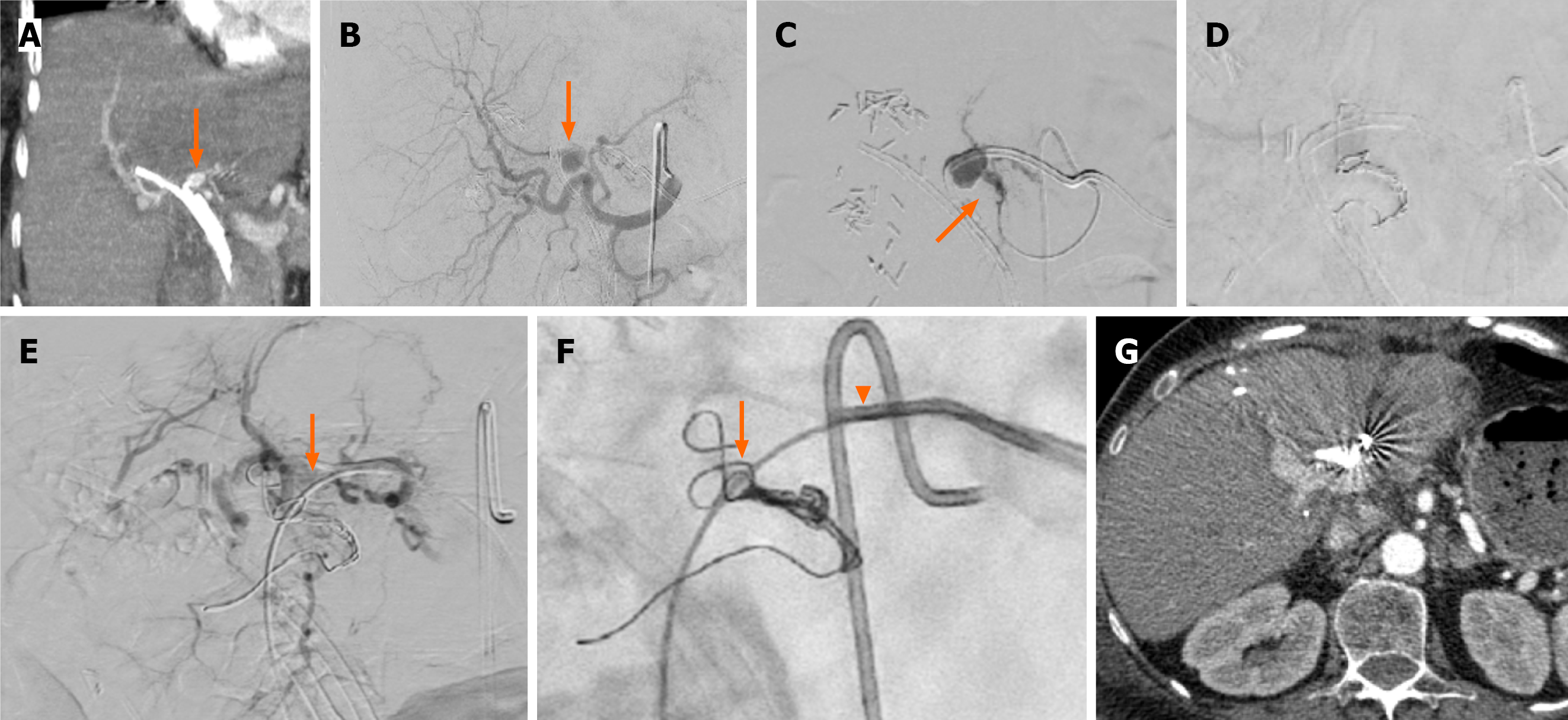
Figure 3 A 53-year-old female patient with colorectal liver hilar metastasis and right lobe endoscopic plastic stent underwent left lobe percutaneous transhepatic biliary drainage.
A: Computed tomography angiography revealed a pseudoaneurysm in proximity to the percutaneous biliary catheter (arrow); B: Selective digital subtraction angiography from the common hepatic artery confirmed the presence of the pseudoaneurysm arising from the left hepatic artery (arrow); C and D: The left hepatic artery was selectively catheterized with a 1.98Fr microcatheter and proximately embolized with pushable microcoils; as the back to front door” technique was not feasible (microcatheter was not advancing distal to the lesion) the catheter was removed over a 0.035’ guide wire (arrow), E and F: An 8Fr sheath was placed and using a 5Fr angled catheter (arrow) and the same microcatheter via the 5Fr catheter (arrowhead), the sack of the pseudoaneurysm was embolized using pushable microcoils (arrow). Note that during removal of the biliary catheter arterial blood was noted, confirming the necessity of further embolization from the biliary access; G: Check computed tomography angiography at 7 days, prior to discharge, confirmed the occlusion of the pseudoaneurysm. The patient was discharged asymptomatic.
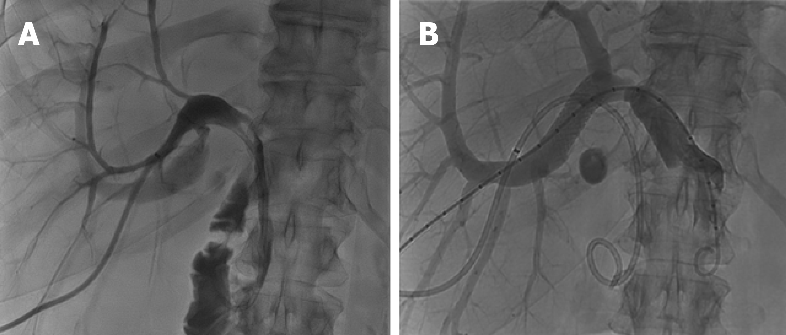
Figure 4 Large perihilar portobiliary fistula in patient with percutaneous transhepatic biliary drainage.
A: Percutaneous transhepatic biliary drainage with evidence of portobiliary fistula; B: A covered stent was inserted in the portal branch to treat the fistula.
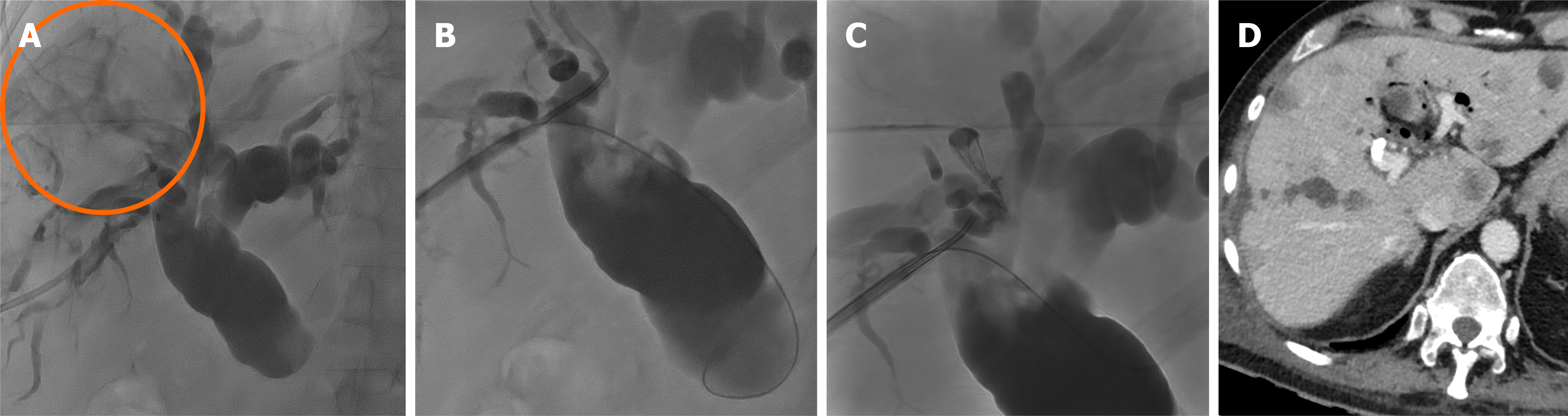
Figure 5 Protobiliary fistula following external biliary drainage insertion.
A and B: Cholangiography via multipurpose catheter shows an iatrogenic bilio-portal fistula (orange circle) occurred during percutaneous transhepatic cholangiography; C: It was treated by N-butyl cyanoacrylate glue embolization of the portal branch; D: Computed tomography scan revealed glue deposition and complete embolization of the fistula.
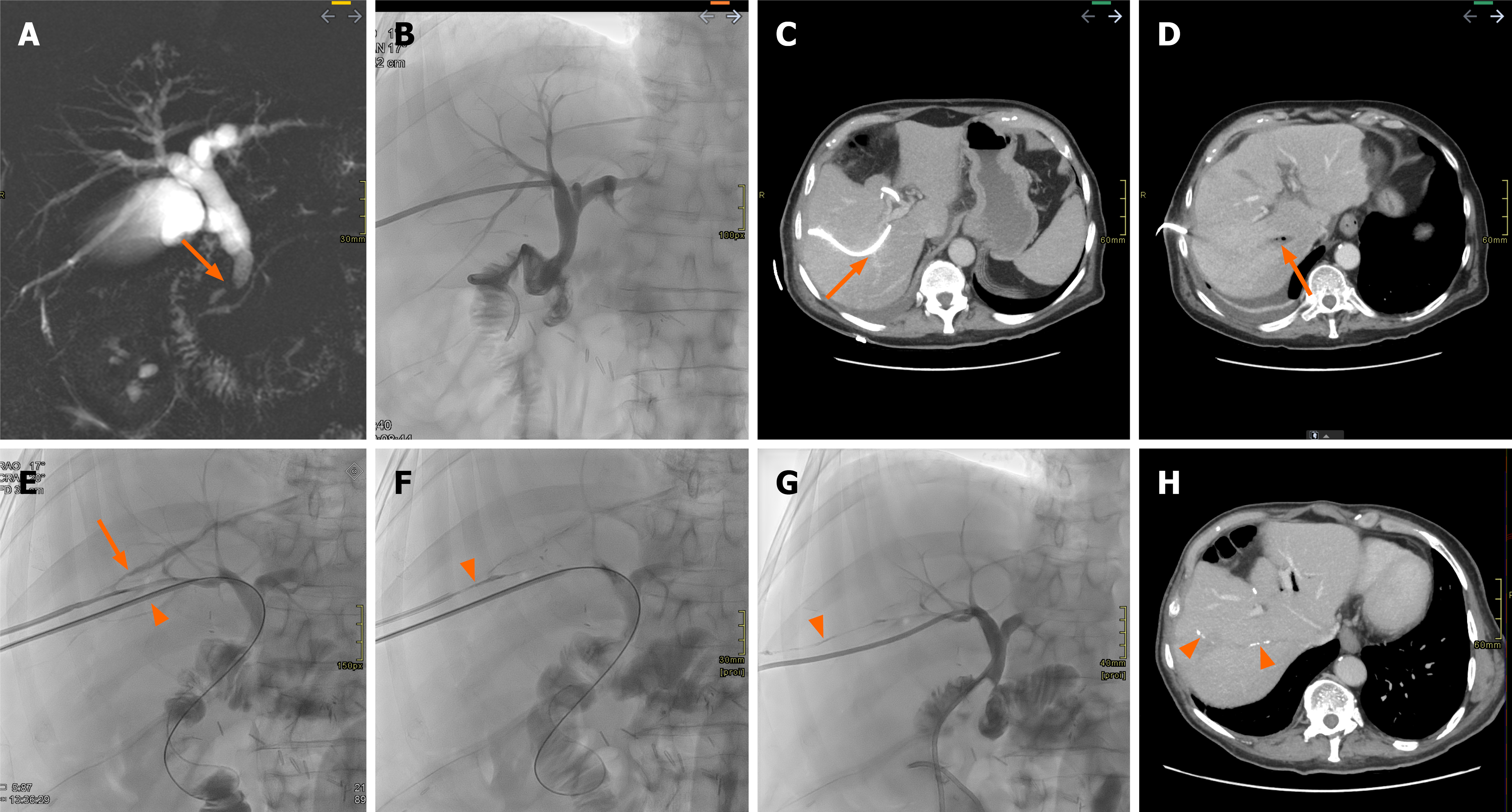
Figure 6 A 73-year-oid patient with cholangiocarcinoma and bile leak.
A: Magnetic resonance cholangiopancreatography demonstrates a filling defect in the distal common bile duct (arrow), later diagnosed as cholangiocarcimona. The patient subsequently underwent surgical resection with biliodigestive anastomosis; B: Bile leakage was observed from the surgical drain, prompting percutaneous transhepatic cholangiography. In the days following the percutaneous transhepatic cholangiography, the patient developed sepsis; C: Computed tomography was performed and revealed a marked angulation of the percutaneous drain in close proximity to vascular structures (arrow); D: Along with thrombosis and the presence of gas bubbles in the right suprahepatic vein (arrow); E: A repeat interventional radiology procedure was carried out, which confirmed the presence of a biliovenous leak (arrow) and a new drain was placed in a different right bile duct (arrowhead); F: Subsequently, the biliovenous fistula tract was embolized using N-butyl cyanoacrylate glue (arrowhead); G: Finally, the previously positioned access route was removed, with continued embolization of the entire access tract (arrowhead); H: On follow-up computed tomography after removal of the biliary drain and resolution of the patient’s clinical condition, faint remnants of the embolic material are still visible (arrowheads).
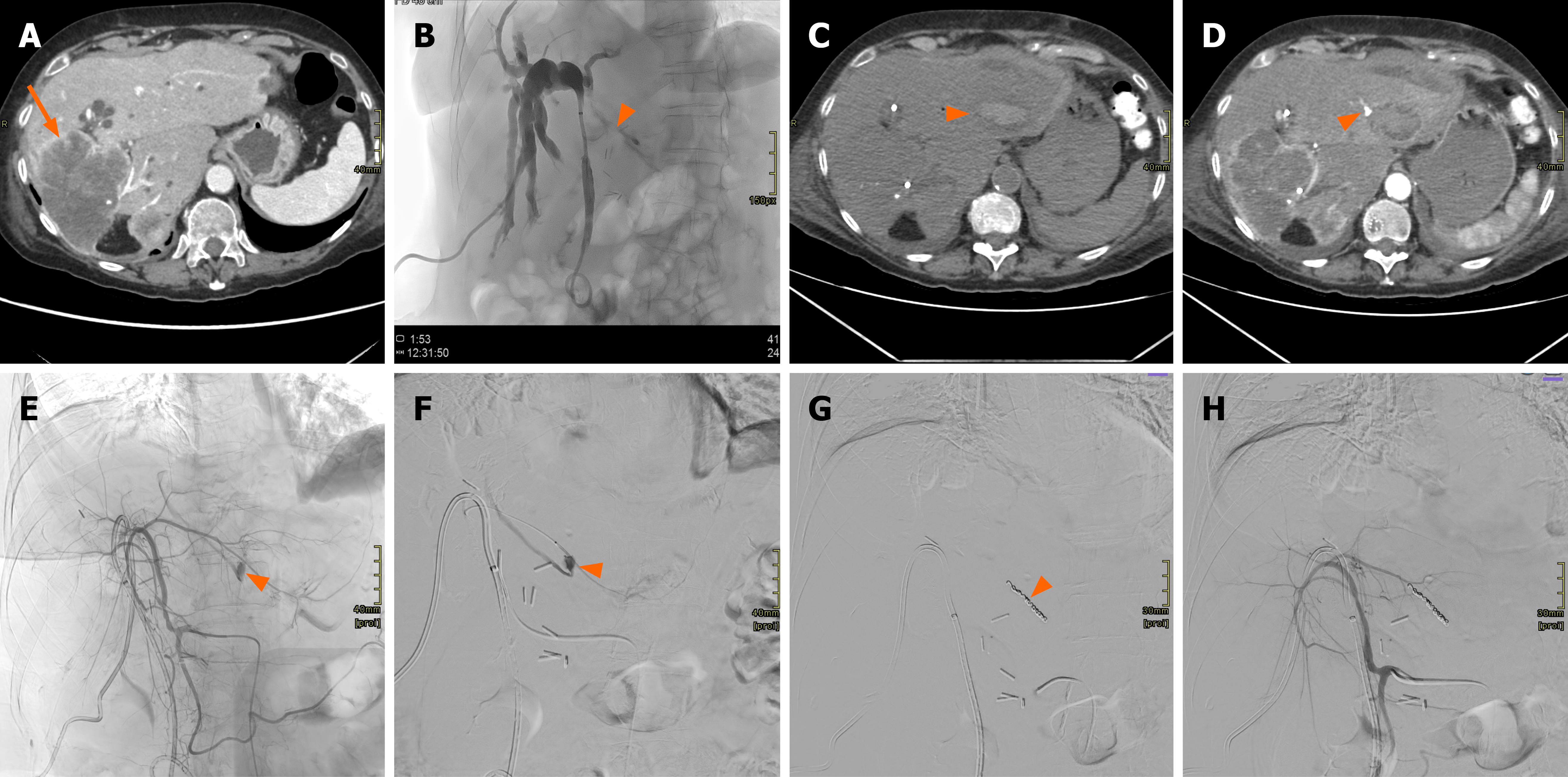
Figure 7 Patient with recurrent cholangiocarcinoma involving segment IV, approximately 18 months after prior right hepatectomy.
A: Computed tomography scan showing a large mass (arrow) in the right liver lobe, in keeping with cholangiocarcinoma; B: The patient underwent percutaneous transhepatic cholangiography for hyperbilirubinemia. An initial attempt at percutaneous biliary access to segment III was unsuccessful; residual contrast medium can be seen in segment III (arrowhead); C: Due to the onset of severe epigastric pain, computed tomography was performed, revealing a large hematoma in the left hepatic lobe (arrowhead); D: With evidence of arterial blush from a branch supplying segment III (arrowhead); E: Angiographic evaluation confirmed this finding, both on common hepatic artery catheterization (arrowhead); F: And during selective angiography (arrowhead); G: Embolization of the arterial lesion was performed using coils (arrowhead); H: The final proximal angiographic control demonstrated successful exclusion of the treated branch from arterial flow, with no further contrast extravasation.
- Citation: Acquafredda F, Vizzuso A, Grigoriadis S, Di Giovanni G, Renzulli M, Giampalma E, Paraskevopoulos I, Spiliopoulos S, Inchingolo R. Vascular complications in percutaneous biliary interventions. World J Radiol 2026; 18(8): 119018
- URL: https://www.wjgnet.com/1949-8470/full/v18/i8/119018.htm
- DOI: https://dx.doi.org/10.4329/wjr.119018