
















Copyright: ©Author(s) 2026.
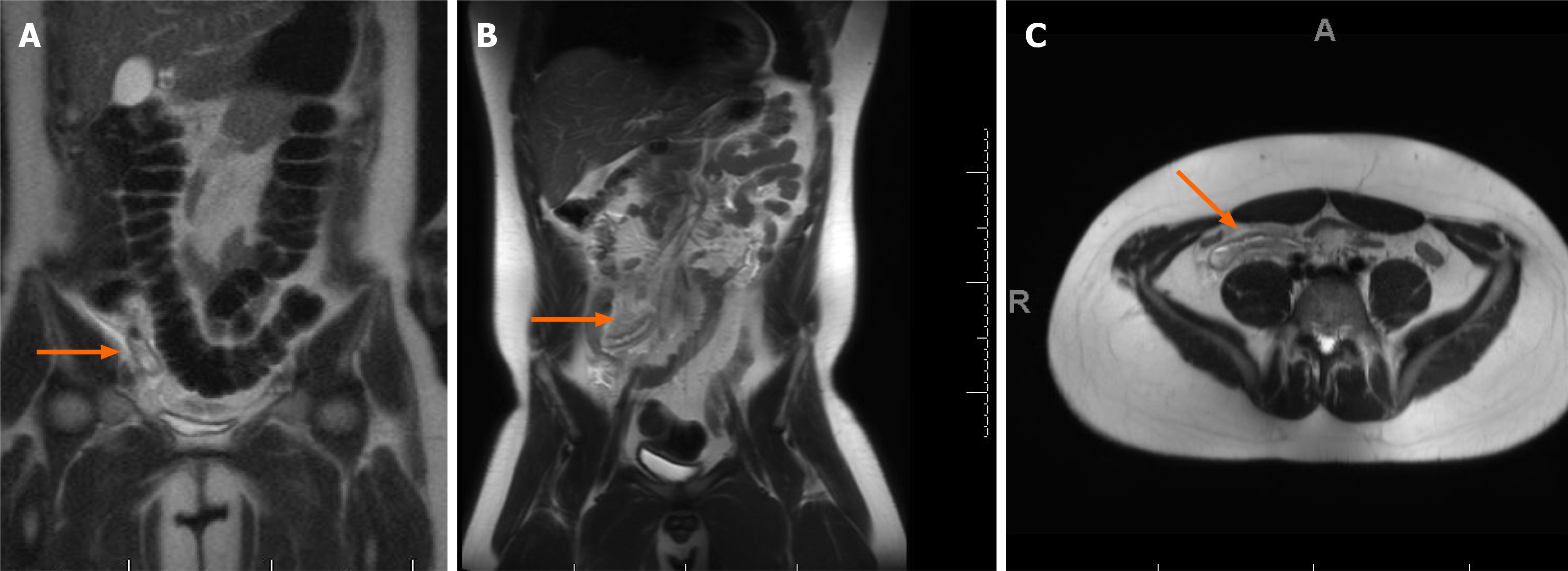
Figure 1 Acute appendicitis on magnetic resonance imaging.
A: Coronal view of adult patient demonstrating a blind ending pouch with peripheral free high signal fluid, consistent with acute appendicitis (orange arrow); B and C: Coronal (B) and axial (C) T2 weighted magnetic resonance imaging views of pediatric patient with a thickened blind ending pouch in the right lower quadrant with surrounding enhancement and fluid, consistent with appendicitis (orange arrow).
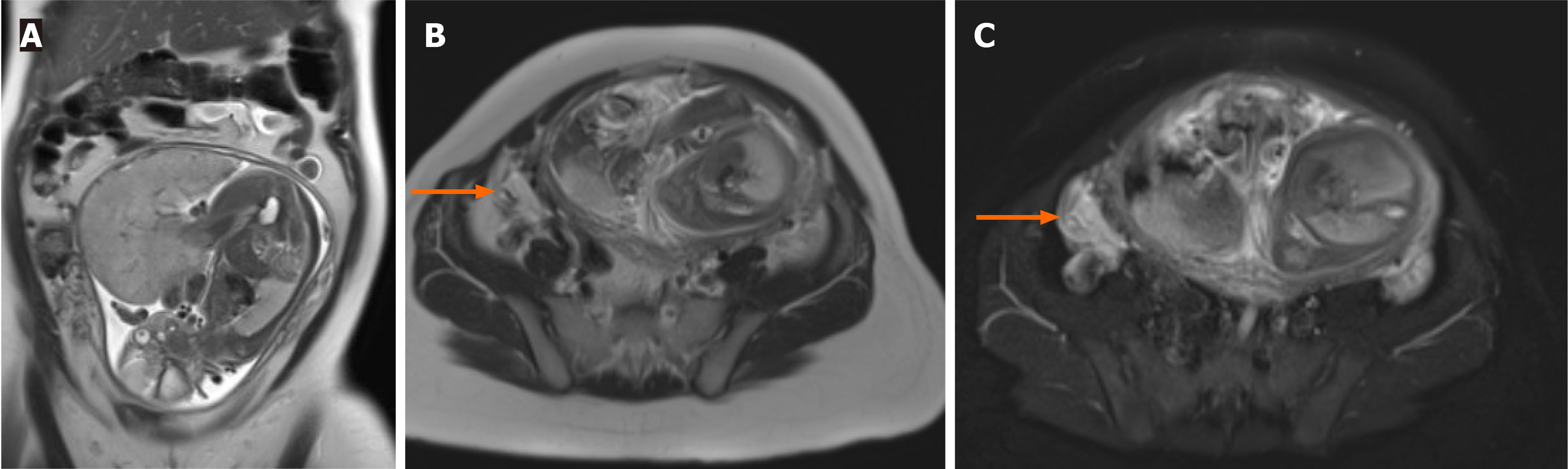
Figure 2 Acute appendicitis in a pregnant patient.
A and B: Coronal (A) and axial (B) T1 weighted images demonstrating a 30-week gestation patient with a thickened appendix and surrounding inflammation, consistent with acute appendicitis; C: Axial T1 fat suppressed weighted image of the same patient highlighting the enhancing fluid in the right lower quadrant surrounding the appendix.
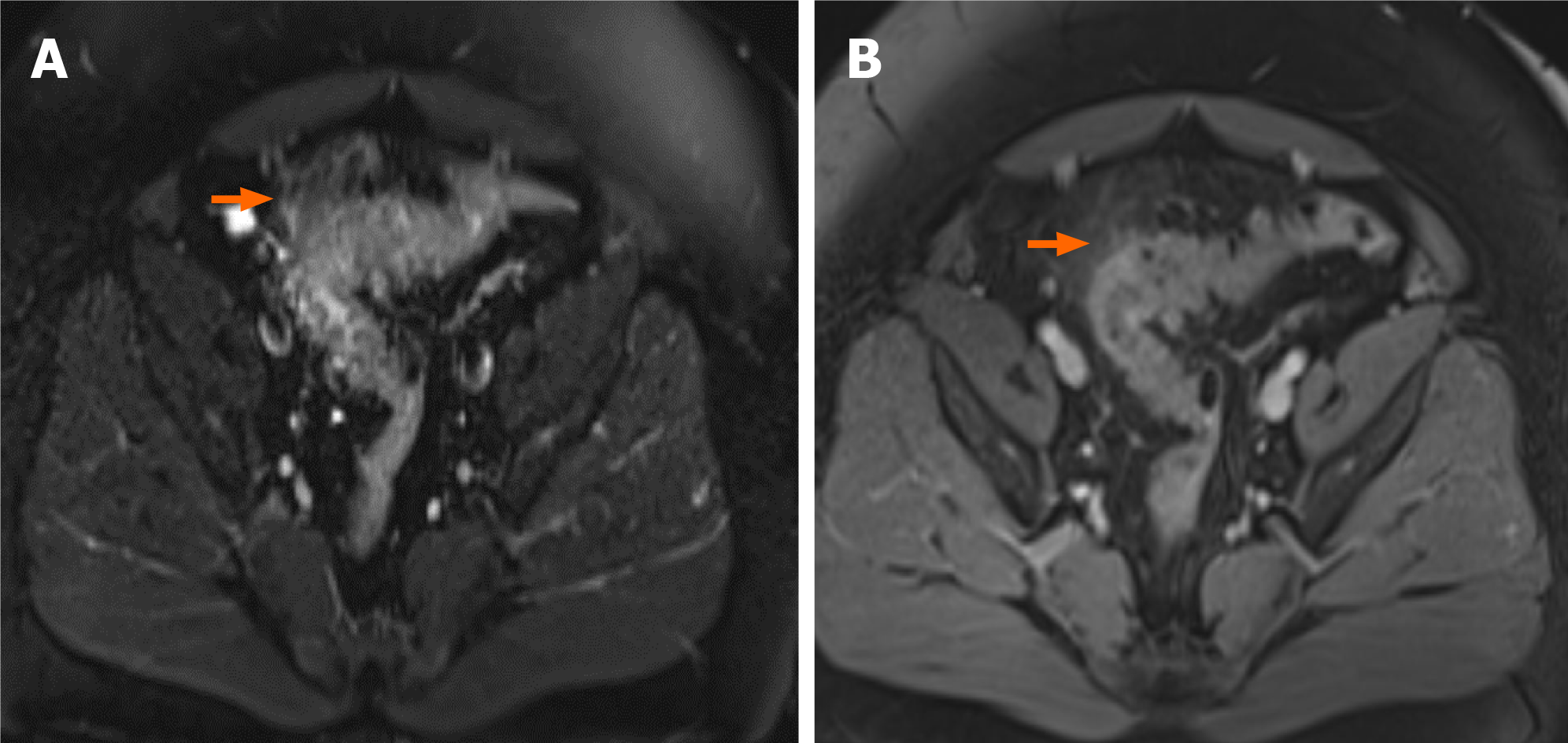
Figure 3 Sigmoid diverticulitis on magnetic resonance imaging.
A: T1-fat suppressed axial magnetic resonance imaging (MRI); B: T2 MRI. Both demonstrate a thickened sigmoid colon with diverticula and surrounding edema (orange arrows). No evidence of rim enhancing fluid collection.
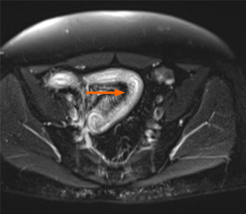
Figure 4 Radiation enteritis.
T1-weighted post-contrast fat-saturated magnetic resonance image highlighting a short segment of the ileum with luminal narrowing (orange arrow), submucosal enhancement and bowel wall thickening following pelvic radiation.
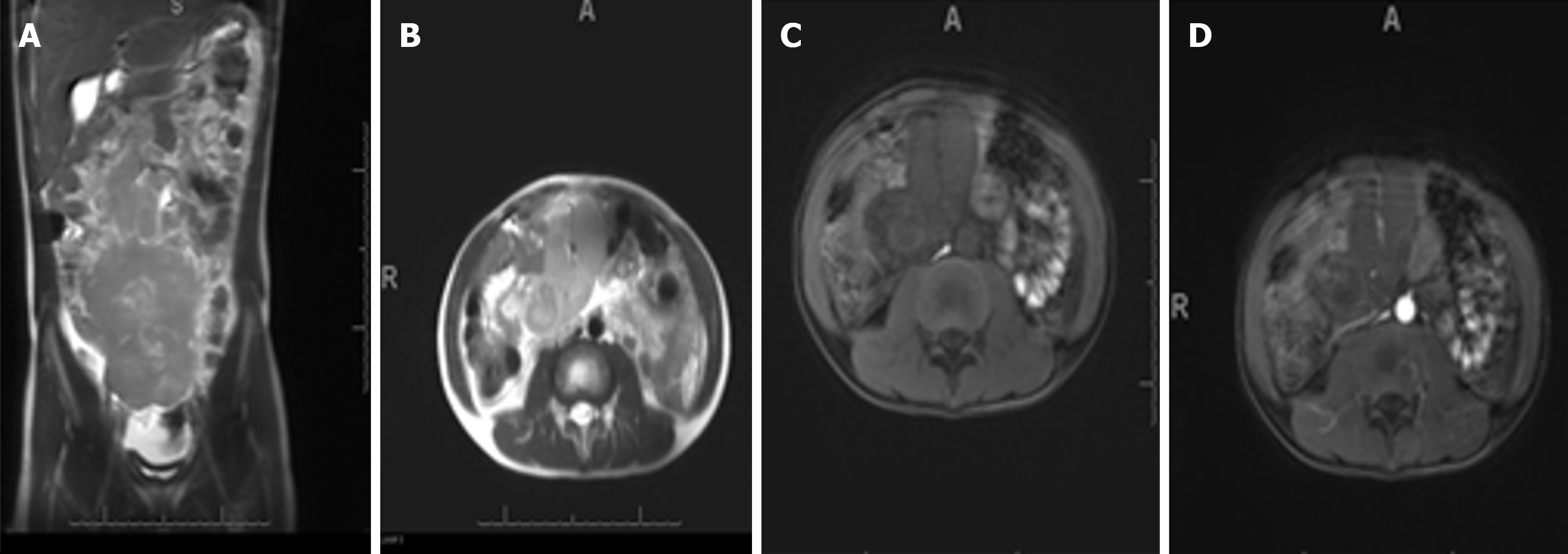
Figure 5 Small bowel lymphoma on magnetic resonance imaging.
A: Coronal T1 image; B: Axial T1 image; C and D: Axial T1-fat suppressed magnetic resonance imaging. All images of the abdomen of a 10-year-old pediatric patient show a homogeneously thickened small bowel loop measuring approximately 5 cm in thickness. This aneurysmal dilation of small bowel within the muscularis propria is consistent with small bowel lymphoma.
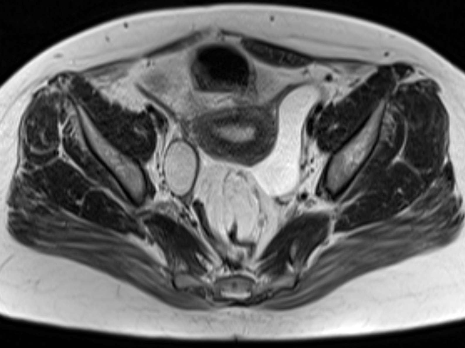
Figure 6 Mild pouchitis on magnetic resonance imaging.
Axial T2 half-Fourier acquisition single-shot turbo spin-echo image of the pelvis in a female patient with a history of total proctocolectomy with ileal anal anastomosis showing a thickened ileal wall with surrounding free fluid, suggestive of mild pouchitis.
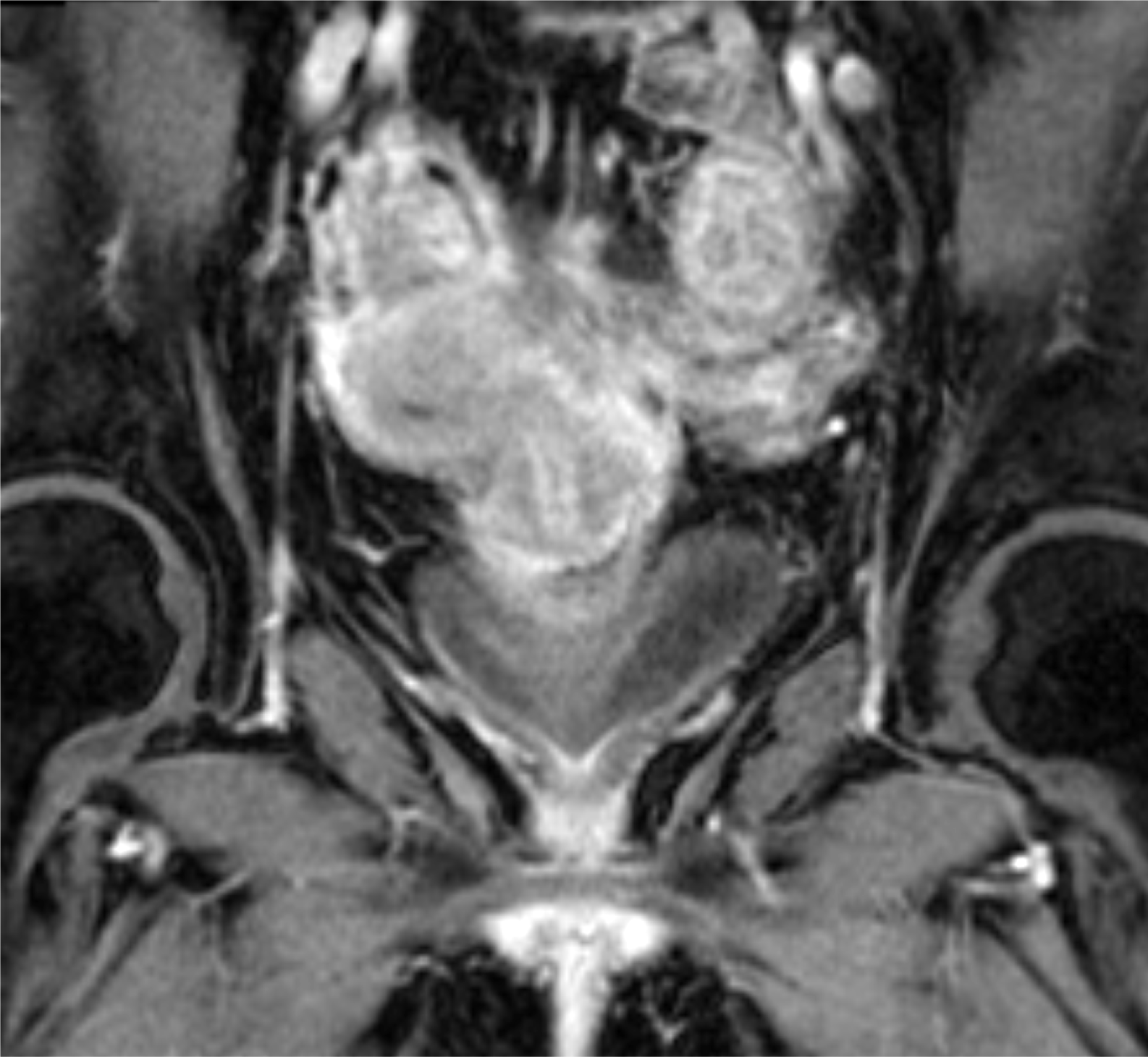
Figure 7 Active pouchitis on magnetic resonance imaging.
Coronal T1 volumetric interpolated breath-hold examination fat-saturated contrast enhanced magnetic resonance imaging showing thickened and edematous submucosa of the J-pouch in a patient with acute flare of known Crohn’s disease, consistent with active pouchitis.
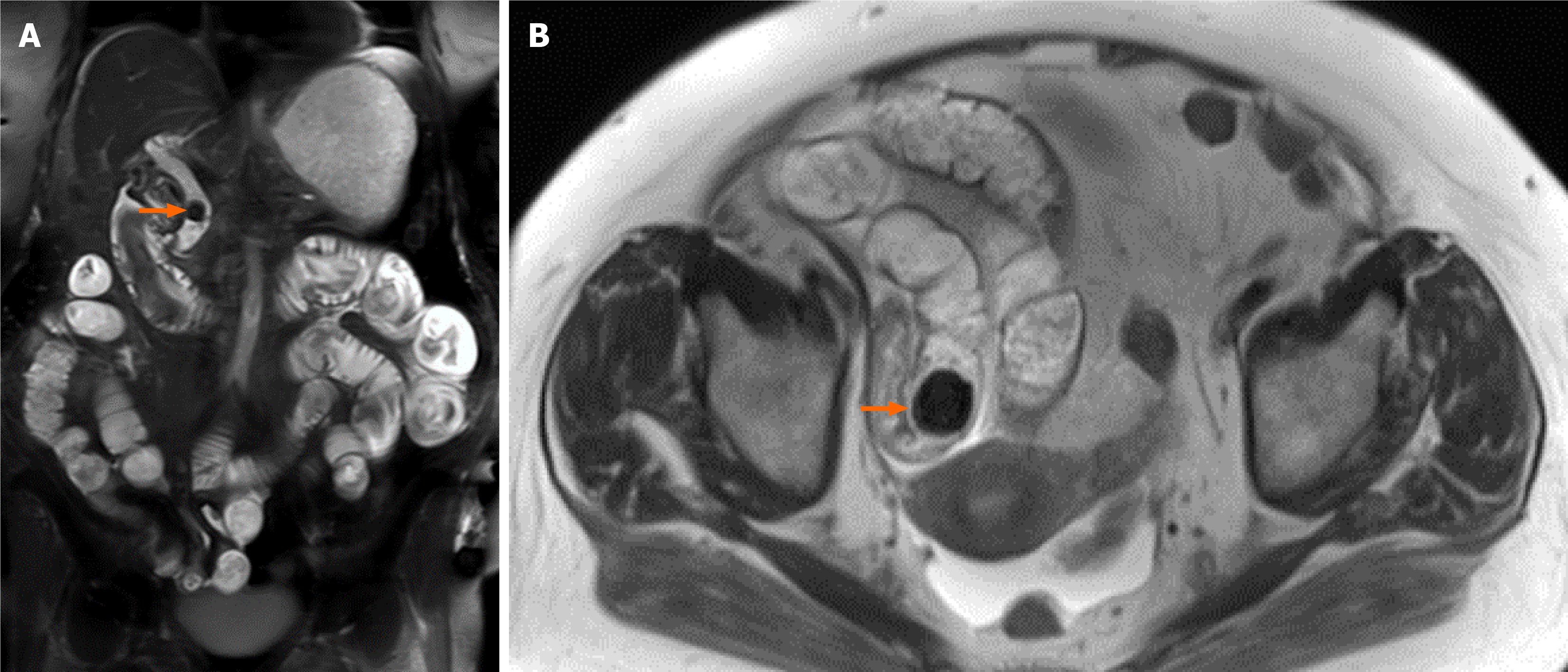
Figure 8 Gallstone ileus on magnetic resonance imaging.
A: Coronal T1 fat suppressed magnetic resonance imaging of the abdomen demonstrating a common bile duct stone (orange arrow) and diffusely distended small bowel loops; B: Axial T2 image showing an additional stone in the distal ileal loop causing proximal obstruction (orange arrow). These findings are in keeping with gallstone ileus.
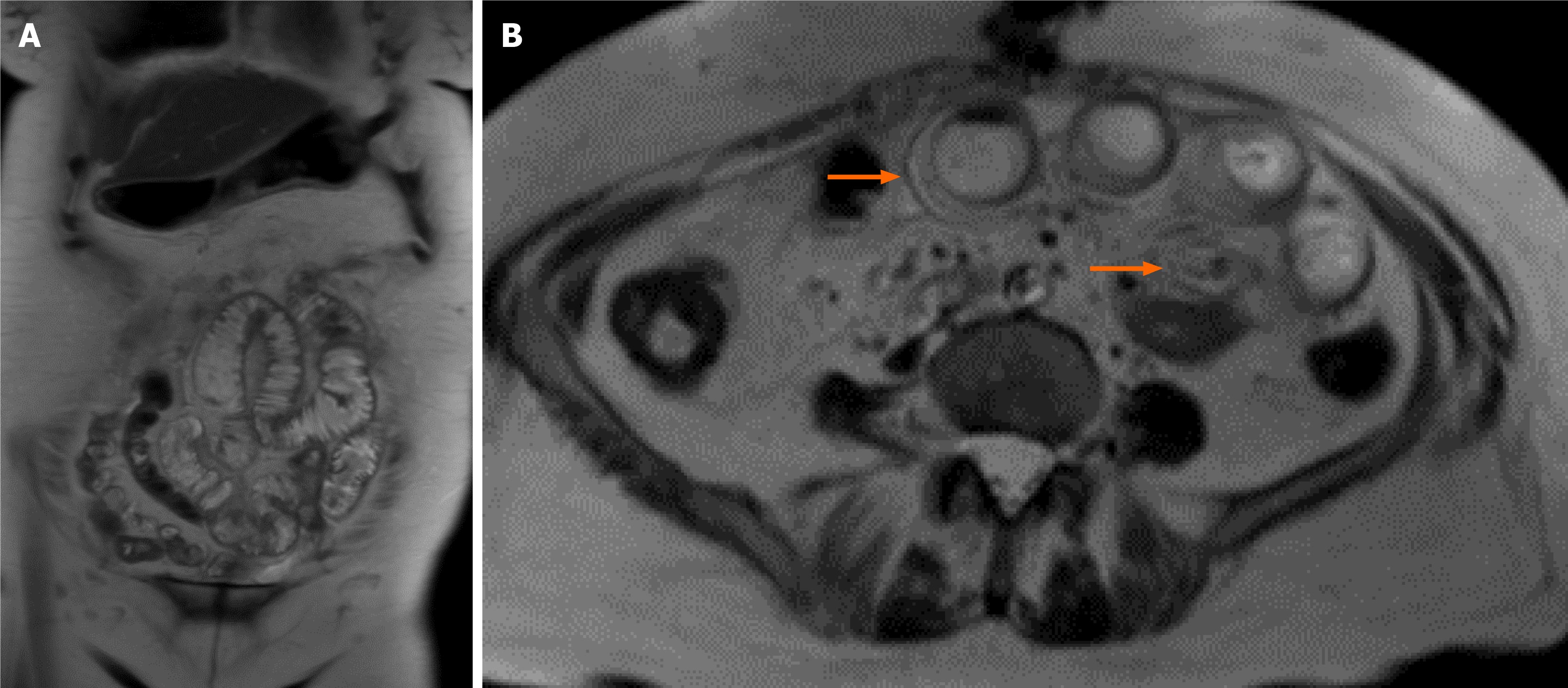
Figure 9 Small bowel obstruction secondary to adhesions.
A and B: Coronal (A) and axial (B) T1-weighted magnetic resonance imaging of the abdomen demonstrating radial orientation of distended small bowel loops with mural enhancement (orange arrows) in an obstruction secondary to adhesions.
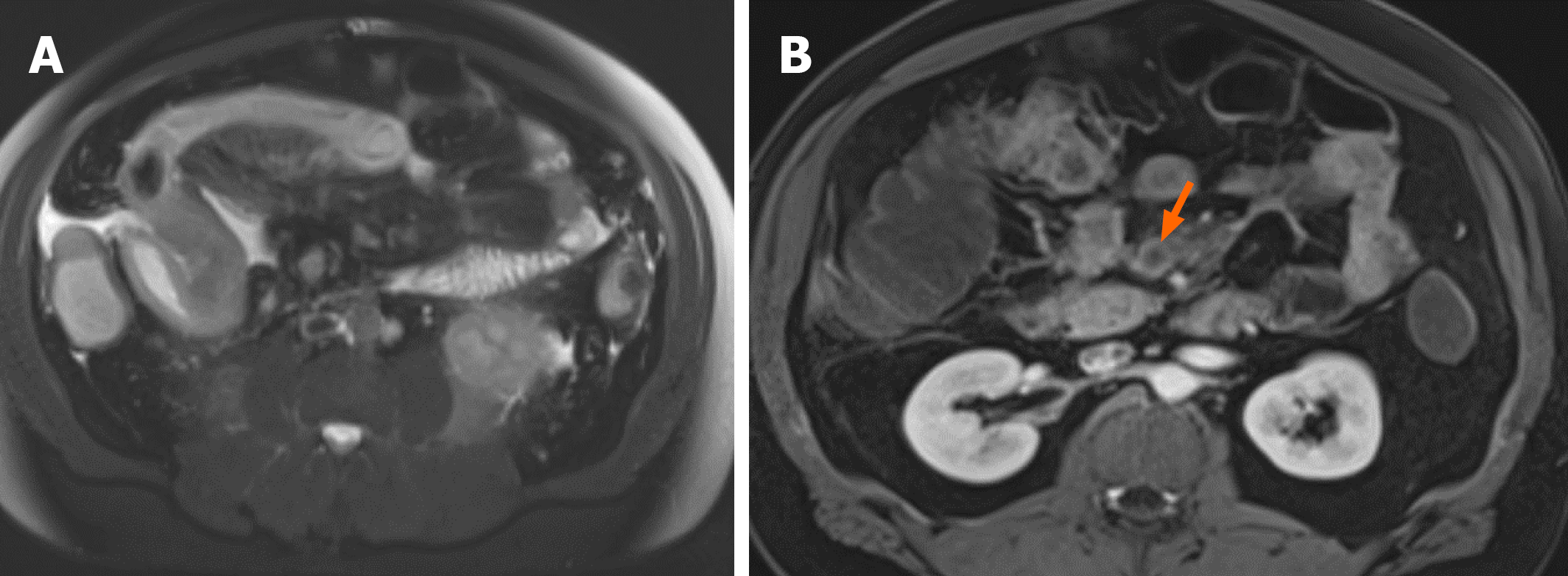
Figure 10 Mesenteric ischemia secondary to superior mesenteric vein thrombosis.
A: T2-weighted fat-saturated axial magnetic resonance imaging of the abdomen demonstrating diffuse ascending and transverse colon wall thickening, submucosal edema and pericolonic free fluid at the level of the hepatic flexure; B: T2-weighted post-contrast fat-saturated axial image demonstrating occlusive thrombus in the superior mesenteric vein (orange arrow). Findings are concordant with wet ischemia secondary to superior mesenteric vein thrombosis.
- Citation: Gonzalez Baerga CI, Menendez Santos M, Ortiz Cordero RG, Guevara Tirado OA, Virarkar M, Gopireddy DR. Role of magnetic resonance in bowel pathologies: Beyond inflammatory bowel disease. World J Radiol 2026; 18(7): 122165
- URL: https://www.wjgnet.com/1949-8470/full/v18/i7/122165.htm
- DOI: https://dx.doi.org/10.4329/wjr.122165