
















Copyright: ©Author(s) 2026.
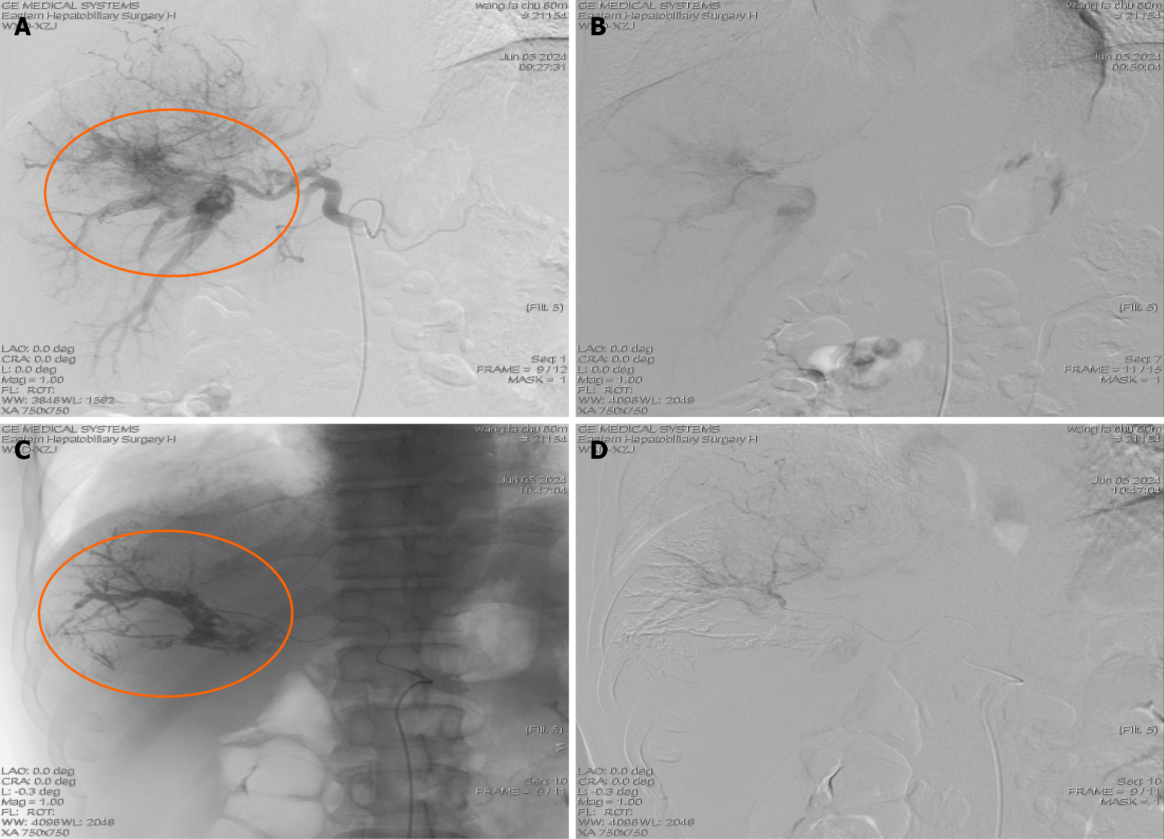
Figure 1 Arterio-portal shunt detection on hepatic arteriography and closure by portal vein embolization.
A: Hepatic arteriography detected Arterio-portal shunt (APS); B: Hepatic right arteriography with microcatheter; C: Embolization of the responsible portal vein with N-butyl cyanoacrylate and iodized oil mixture; D: Repeated hepatic right artery angiography with a microcatheter showed that the APS had been completely blocked.
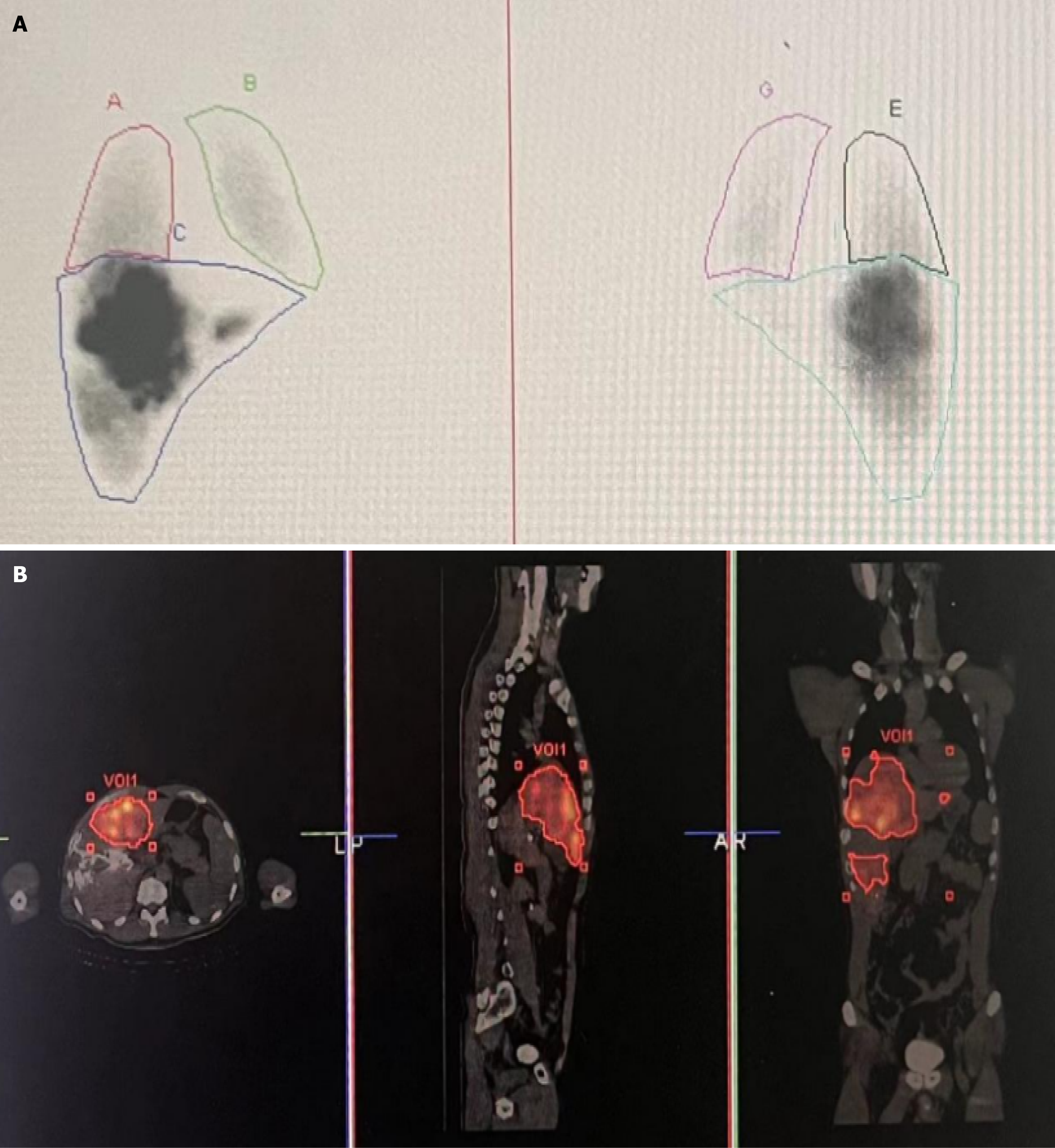
Figure 2 Single photon emission computed tomography/computed tomography scintigraphy following injection of (99m) technetium-labeled macroaggregated albumin ((99m)Tc-MAA) into the tumor-feeding artery.
A: Planar scintigraphy to calculate the hepatopulmonary shunt. B: Transverse, sagittal and coronal single photon emission computed tomography/computed tomography imaging of the distribution of (99m)Tc-MAA in tumor lesions, normal liver tissue and other systemic regions.
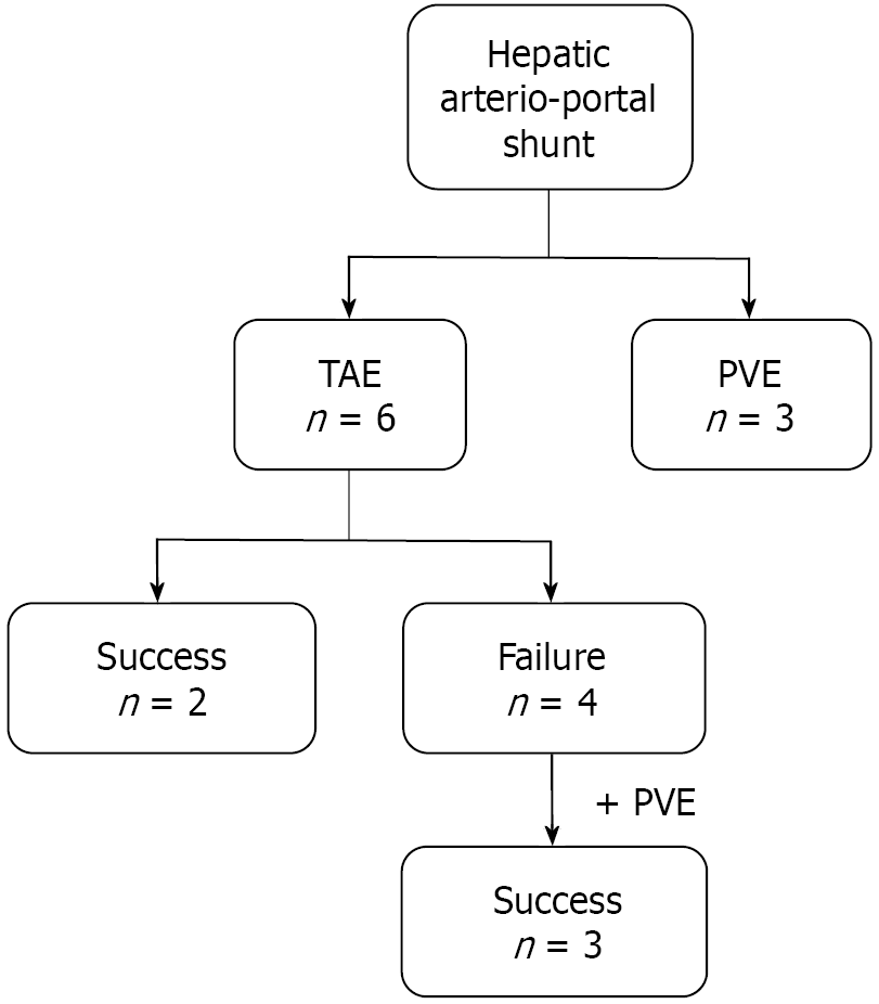
Figure 3 Flowchart of closure of the hepatic arterio-portal shunt in hepatocellular carcinoma patients during the mapping procedure.
TAE: Transcatheter arterial embolization; PVE: Portal vein embolization.
- Citation: Liu X, Zhao JY, Xu ZY, Shen SQ, Xu ZJ, Yang YF, Ge NJ, Wang XD. Portal vein embolization for arterio-portal shunt closure in hepatocellular carcinoma to facilitate selective internal radiation therapy. World J Radiol 2026; 18(7): 121348
- URL: https://www.wjgnet.com/1949-8470/full/v18/i7/121348.htm
- DOI: https://dx.doi.org/10.4329/wjr.121348