
















Copyright: ©Author(s) 2026.
World J Radiol. May 28, 2026; 18(5): 119223
Published online May 28, 2026. doi: 10.4329/wjr.v18.i5.119223
Published online May 28, 2026. doi: 10.4329/wjr.v18.i5.119223
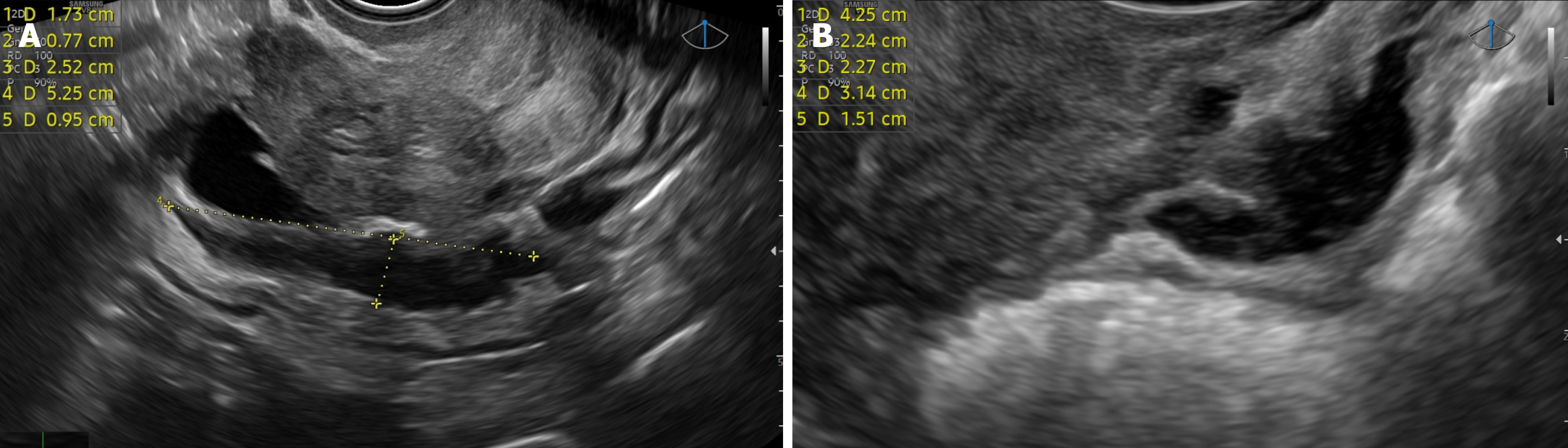
Figure 1 Transvaginal ultrasound findings of endometriotic rectal nodules.
A: Transvaginal ultrasound of an endometriotic rectal nodule; B: Another case of an endometriotic rectal nodule as observed by transvaginal ultrasound.
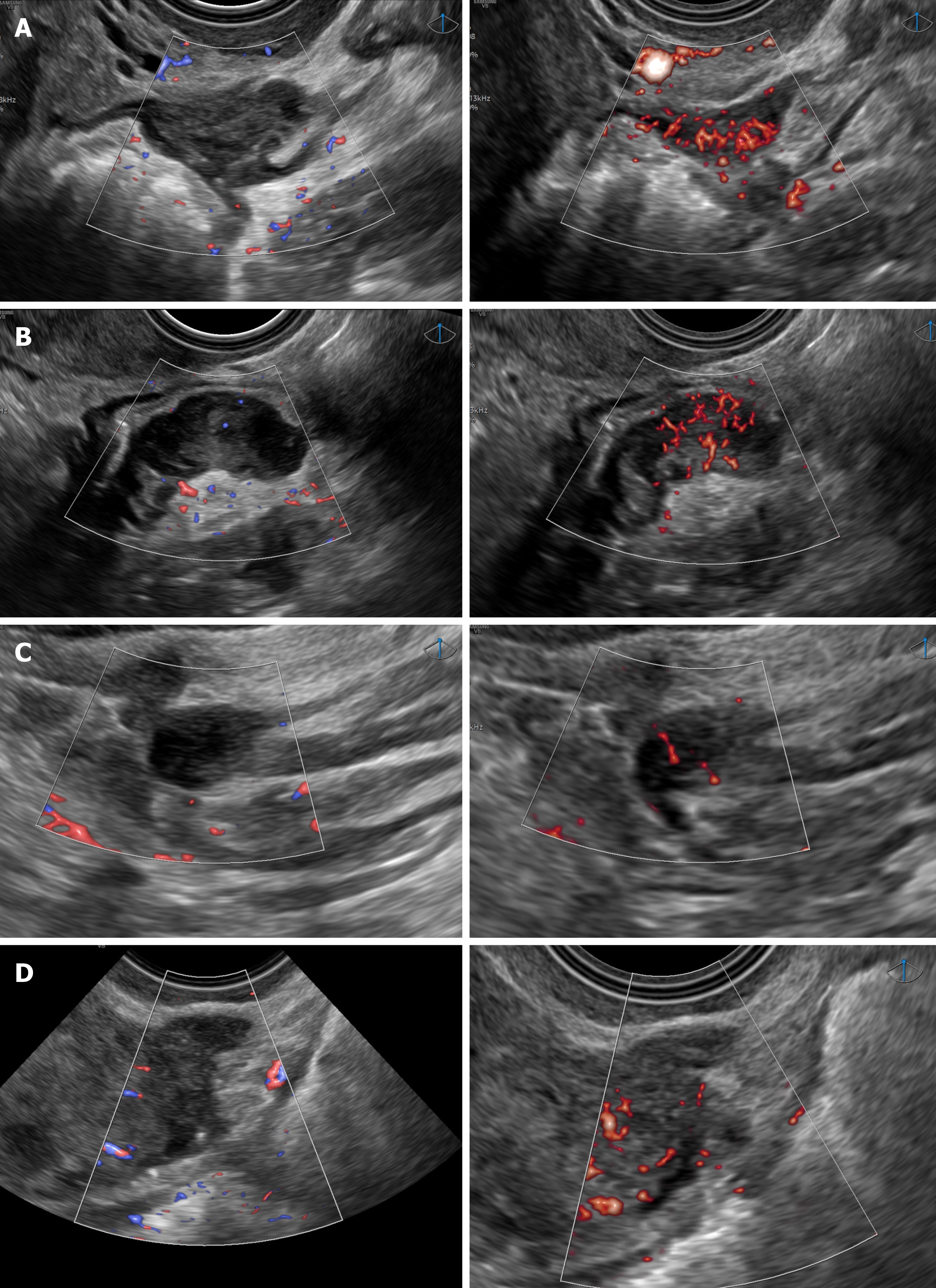
Figure 2 Comparative assessment of rectal nodule vascularization using conventional power doppler and microvascular flow in four cases.
A: Case 1: Power Doppler (vascular score 1) vs microvascular flow (vascular score 4); B: Case 2: Power Doppler (vascular score 1) vs microvascular flow (vascular score 4); C: Case 3: Power Doppler (vascular score 1) vs microvascular flow (vascular score 2); D: Case 4: Power Doppler (vascular score 1) vs microvascular flow (vascular score 2).
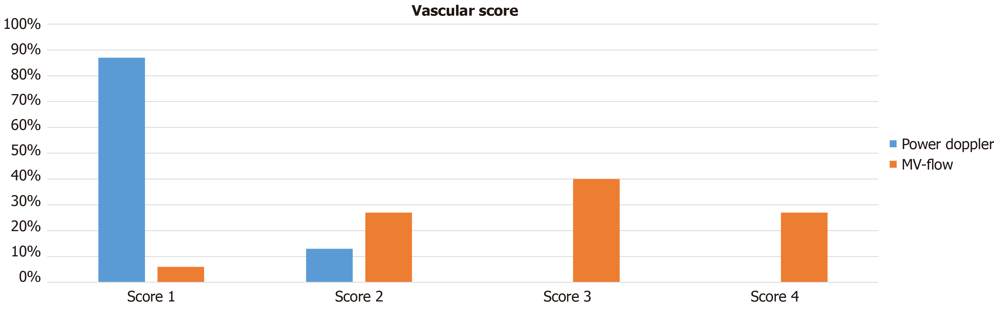
Figure 3 Histogram depicting the distribution of cases according to vascular score using conventional power Doppler and microvascular flow.
MV: Microvascular.
- Citation: Alcazar JL, Pich Barroso DM, Sanchez O, Brunel I, Vilches JC, Orozco R. Enhanced detection of vascularization in rectal endometriosis: A comparative study of microvascular flow and power Doppler. World J Radiol 2026; 18(5): 119223
- URL: https://www.wjgnet.com/1949-8470/full/v18/i5/119223.htm
- DOI: https://dx.doi.org/10.4329/wjr.v18.i5.119223