
















Copyright: ©Author(s) 2026.
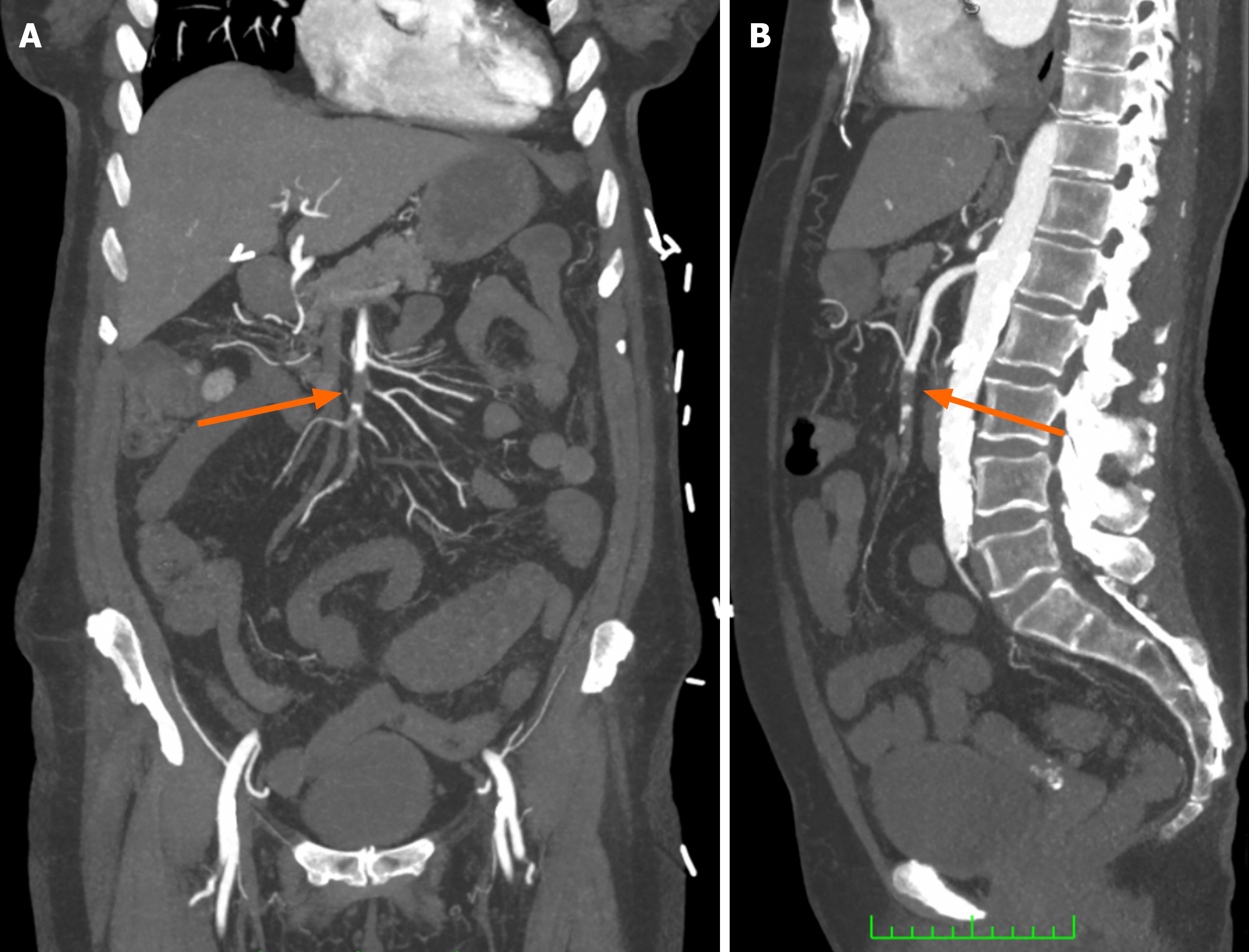
Figure 1 Acute superior mesenteric artery occlusion on computed tomography angiography.
A: Coronal arterial phase image demonstrating an extensive intraluminal filling defect within the middle segment of the superior mesenteric artery (arrow); B: Sagittal arterial phase image demonstrating the same filling defect (arrow). Images from a 65-year-old female presenting with acute embolic mesenteric ischaemia; same patient as Figures 2, 3, and 4.
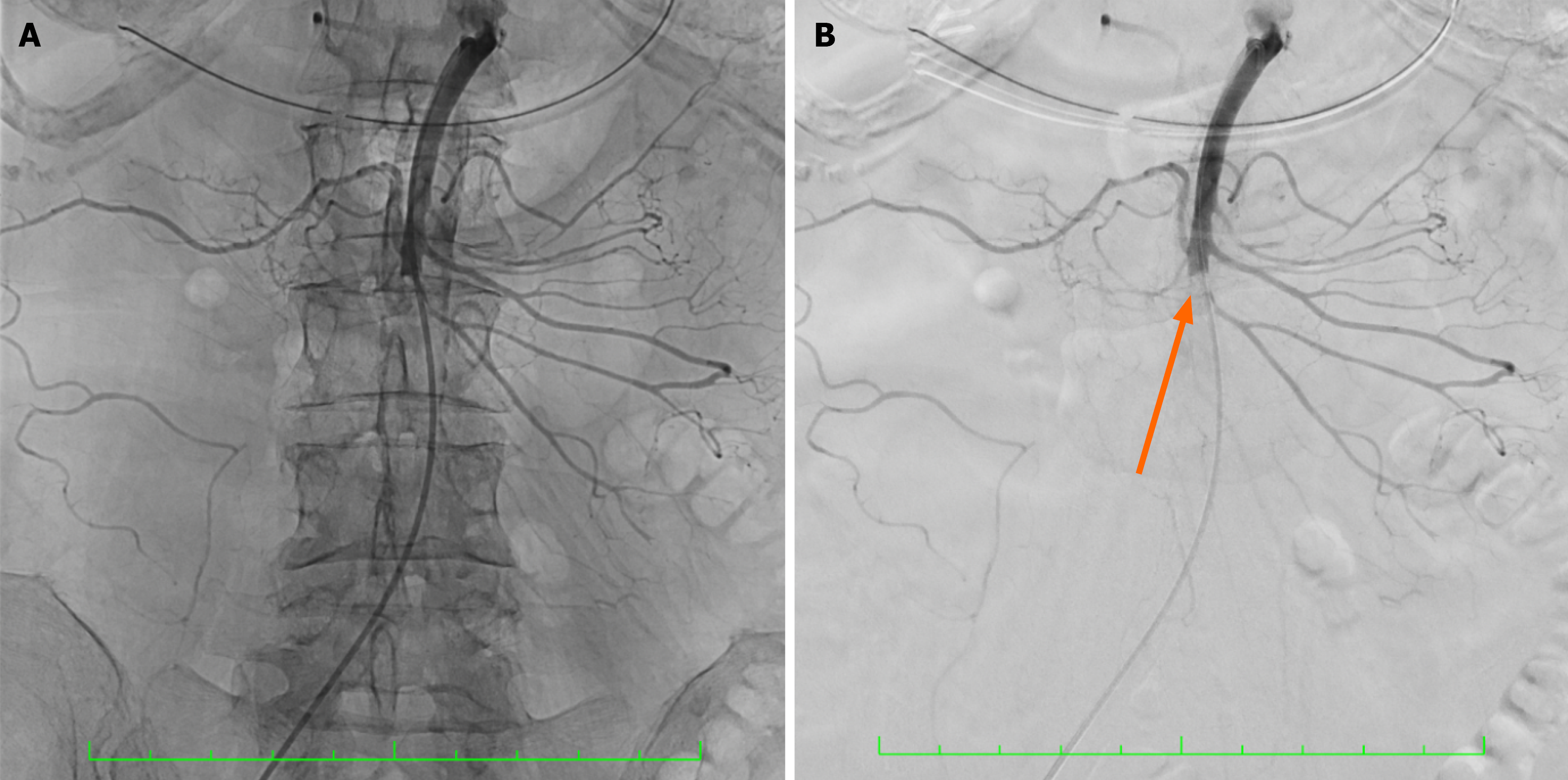
Figure 2 Fluoroscopic and digital subtraction angiographic images of the superior mesenteric artery.
A: Fluoroscopic image during selective superior mesenteric artery angiography; B: Corresponding digital subtraction angiographic image from the same acquisition, demonstrating complete occlusion of the middle segment of the superior mesenteric artery immediately distal to the origins of the right and middle colic arteries (arrow), with no flow in the ileal arcades or ileocolic artery. Same patient as Figures 1, 3, and 4.
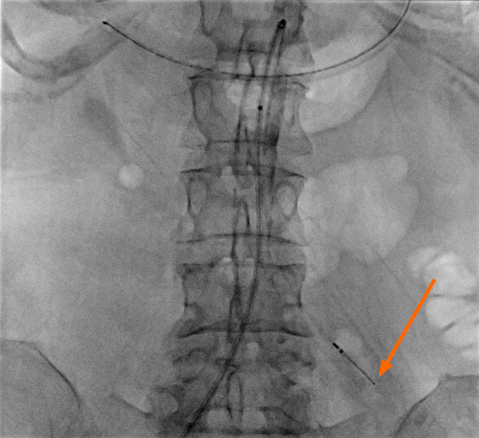
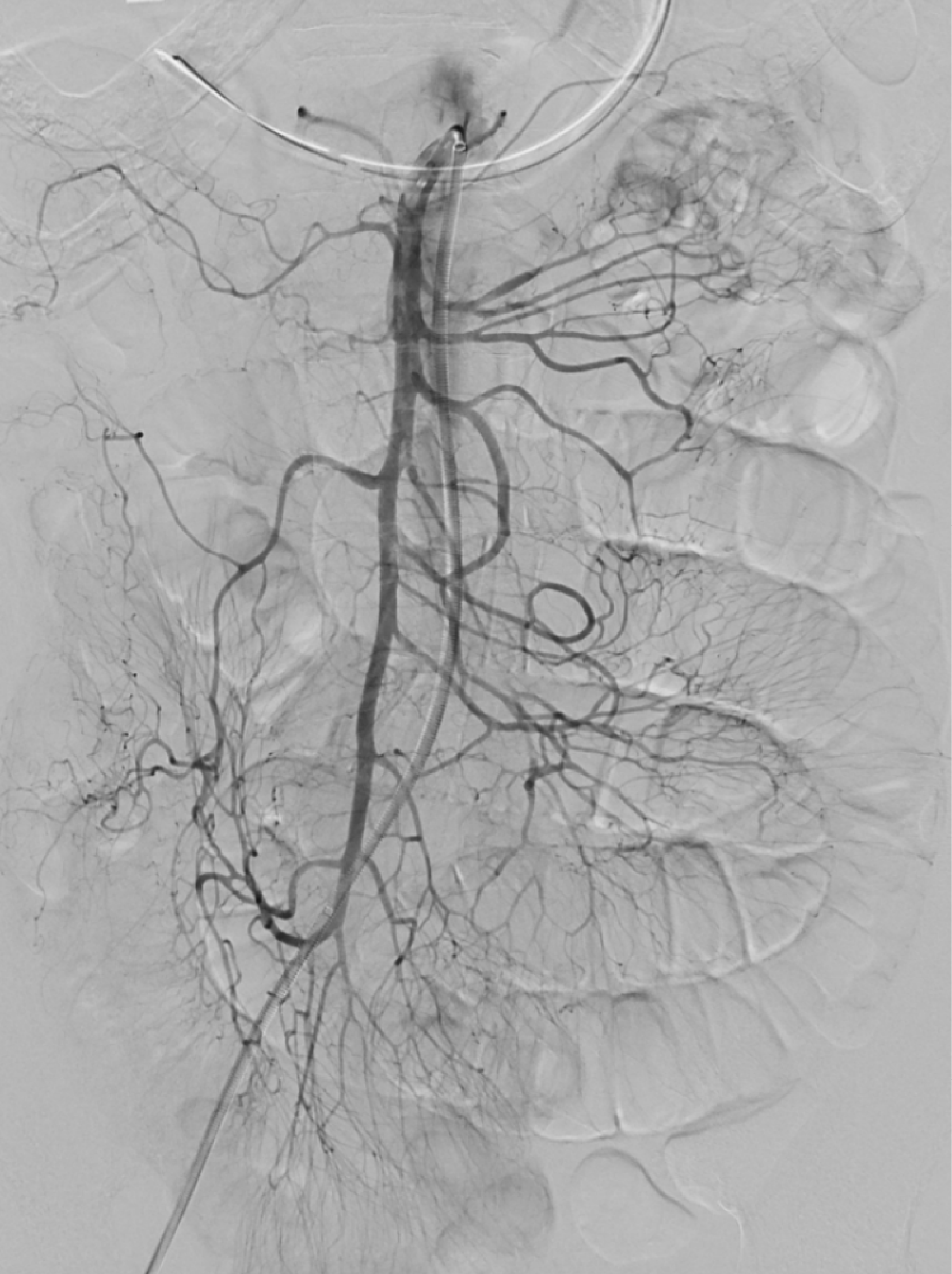
Figure 4 Digital subtraction angiography following catheter-directed thrombolysis.
Anteroposterior digital subtraction angiographic image obtained 24 hours after commencement of catheter-directed thrombolysis, demonstrating complete dissolution of the superior mesenteric artery thrombus with normal contrast opacification of the previously occluded ileal arcades and ileocolic artery. Same patient as Figures 1, 2, and 3.
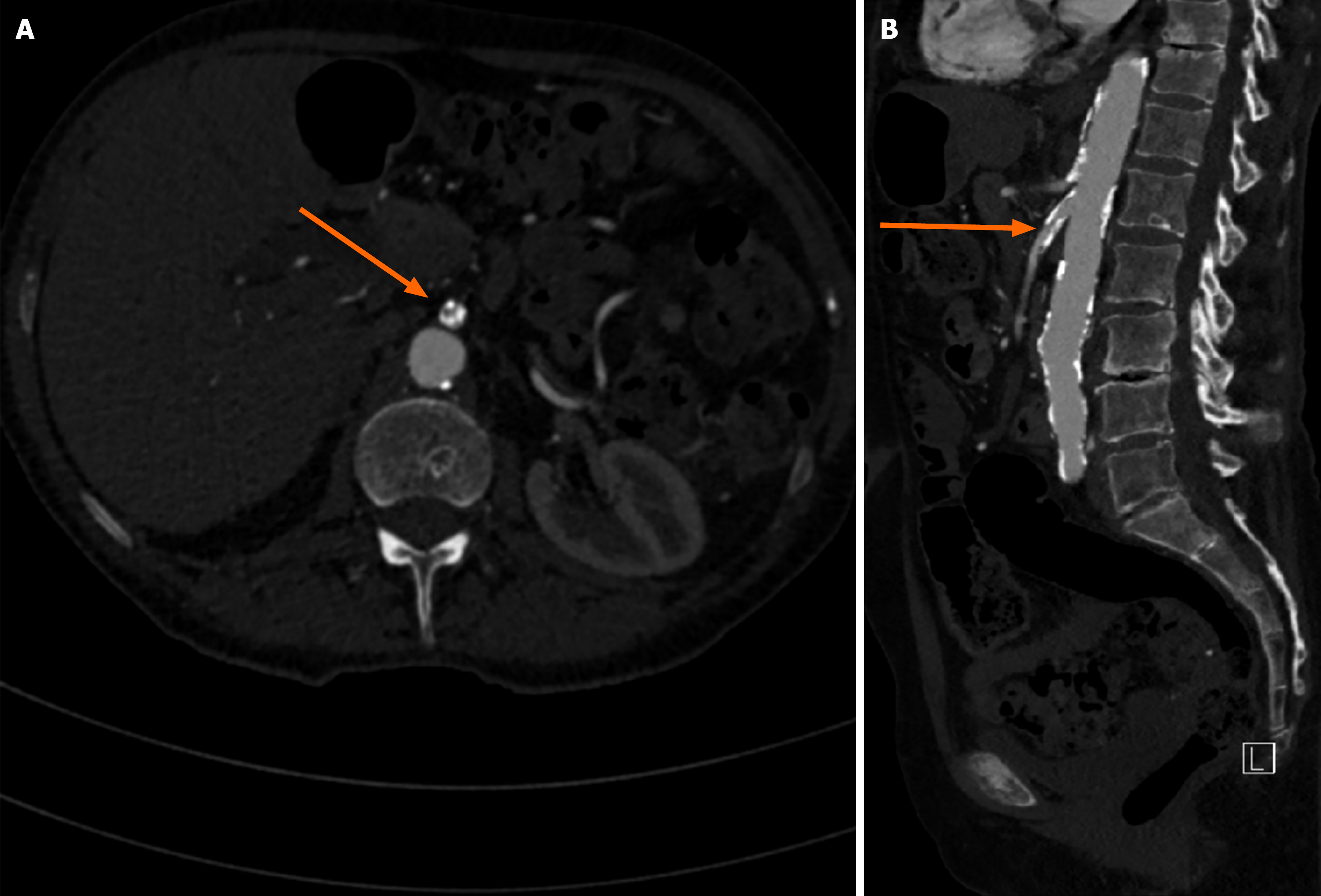
Figure 5 Calcified proximal superior mesenteric artery stenosis on computed tomography angiography.
A: Axial arterial phase image demonstrating heavily calcified atherosclerotic plaque at the proximal superior mesenteric artery (arrow); B: Sagittal arterial phase image demonstrating severe (> 70%) proximal superior mesenteric artery stenosis (arrow). Images from a 76-year-old female with symptomatic chronic mesenteric ischaemia; same patient as Figures 6 and 7.
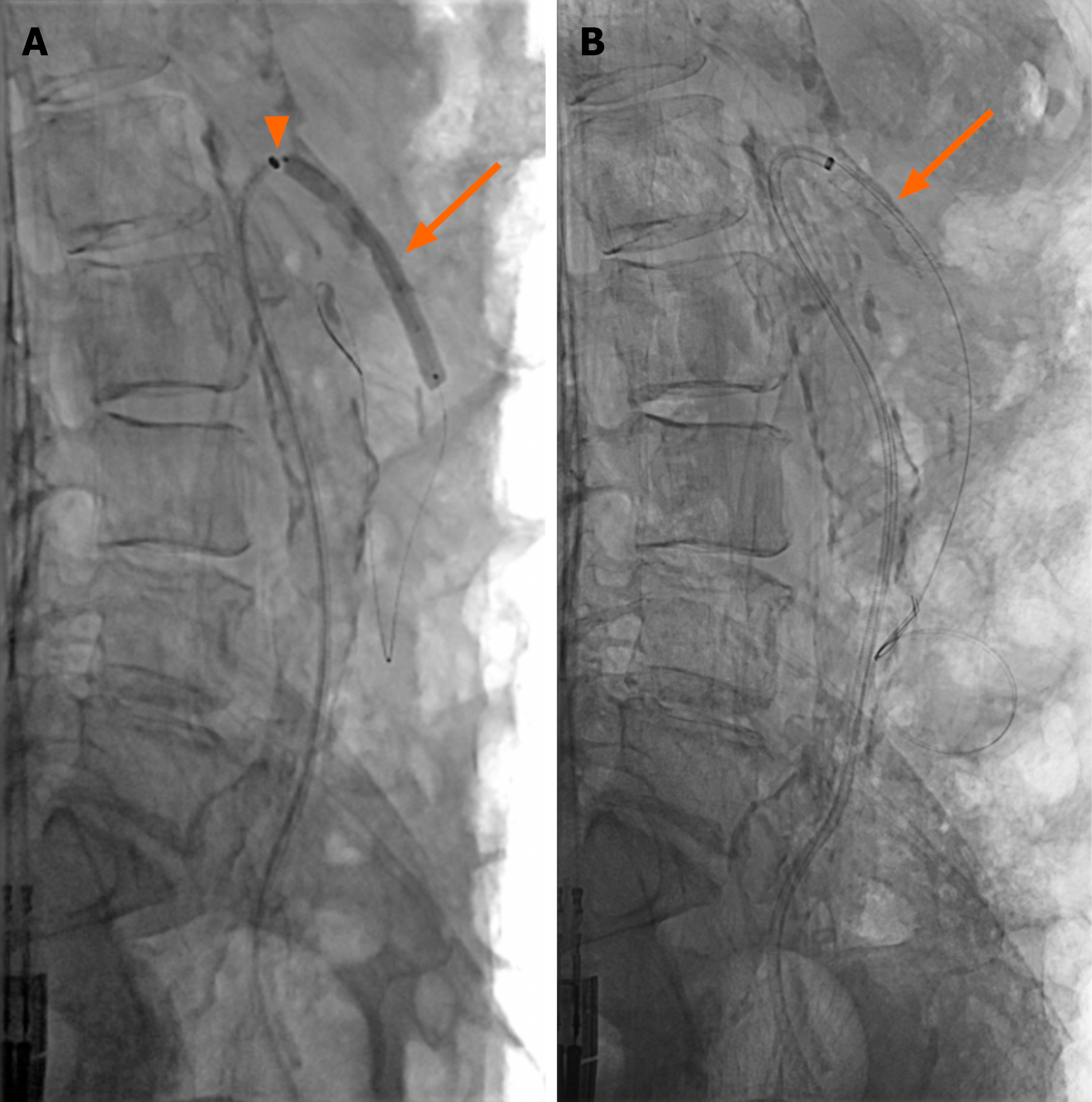
Figure 6 Angioplasty and covered stent placement for proximal superior mesenteric artery stenosis.
A: Lateral fluoroscopic image demonstrating a 45 cm 6-Fr vascular sheath with its tip in the superior mesenteric artery (arrowhead), a 0.018″ guidewire within a distal superior mesenteric artery branch, and balloon angioplasty of the proximal stenosis with a 6 mm balloon (arrow); B: Lateral fluoroscopic image following deployment of a 6 mm covered stent across the proximal stenosis (arrow). Same patient as Figures 5 and 7.
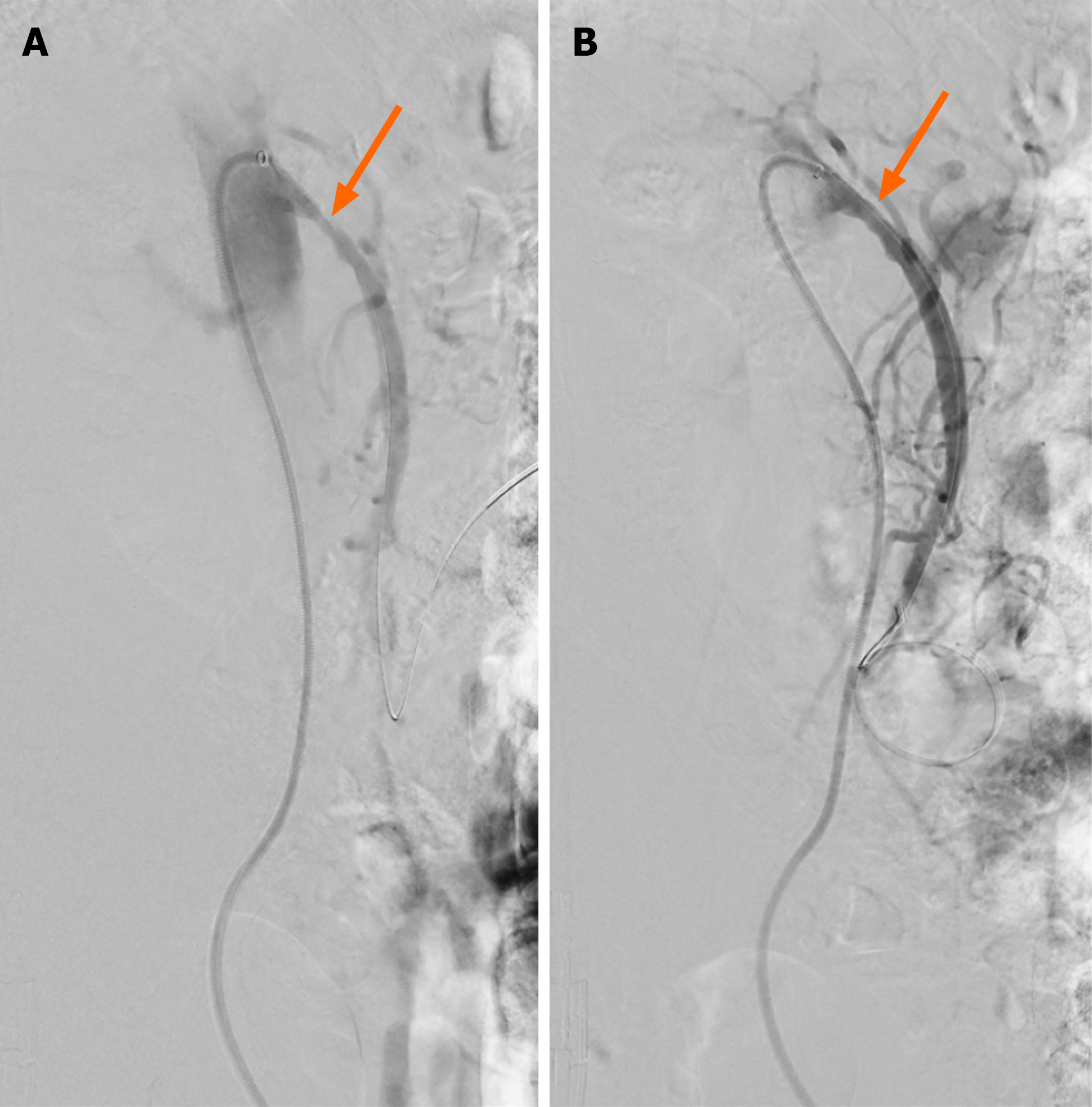
Figure 7 Angiographic result of covered stent placement for proximal superior mesenteric artery stenosis.
A: Lateral digital subtraction angiographic image via a 6-Fr sheath at the ostium of the superior mesenteric artery demonstrating severe proximal stenosis (arrow); B: Corresponding image following covered stent placement demonstrating improved flow across the treated segment, with residual stenosis of approximately 20% secondary to extensive calcified atherosclerosis (arrow). Same patient as Figures 5 and 6.
- Citation: Keady C, O’Malley CM, Mohamed SA, Bell J, Crotty C, Patton A, O’Regan D, Keeling A, McGrath A, Given M, Mulholland D, Sheehan M. Endovascular management of acute and chronic arterial mesenteric ischaemia: A narrative review of evidence informing interventional radiology treatment. World J Radiol 2026; 18(10): 125052
- URL: https://www.wjgnet.com/1949-8470/full/v18/i10/125052.htm
- DOI: https://dx.doi.org/10.4329/wjr.125052