Copyright
©The Author(s) 2025.
World J Radiol. Jul 28, 2025; 17(7): 110385
Published online Jul 28, 2025. doi: 10.4329/wjr.v17.i7.110385
Published online Jul 28, 2025. doi: 10.4329/wjr.v17.i7.110385
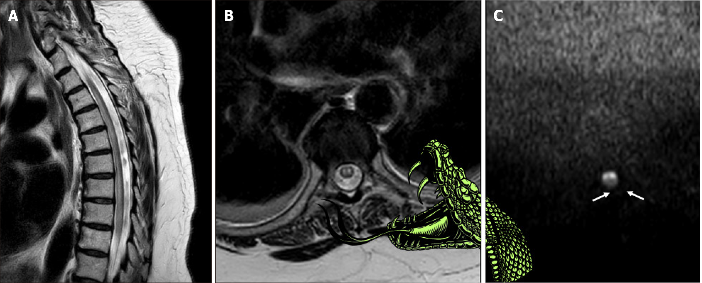
Figure 1 Spinal cord ischemia: “snake bite sign”.
A: Sagittal T2-weighted magnetic resonance image demonstrates longitudinally extensive hyperintensity involving predominantly the anterior and central portions of the thoracic spinal cord; B: Axial T2-weighted magnetic resonance image reveals bilaterally symmetrical punctate areas of high T2 signal intensity (arrows), located at the expected positions of the anterior horn cells. This characteristic appearance resembles puncture wounds from snake fangs, supporting the analogy of the proposed “snake bite sign”, as depicted in the illustration; C: Axial diffusion-weighted magnetic resonance image confirms restricted diffusion (arrows) in the corresponding affected regions, consistent with spinal cord ischemia in the distribution territory of the anterior spinal artery.
- Citation: Arkoudis NA, Karachaliou A, Triantafyllou G, Papadopoulos A, Koutserimpas C, Velonakis G. Spinal cord ischemia: The “snake bite sign”. World J Radiol 2025; 17(7): 110385
- URL: https://www.wjgnet.com/1949-8470/full/v17/i7/110385.htm
- DOI: https://dx.doi.org/10.4329/wjr.v17.i7.110385


