Copyright
©The Author(s) 2025.
World J Radiol. Jul 28, 2025; 17(7): 107140
Published online Jul 28, 2025. doi: 10.4329/wjr.v17.i7.107140
Published online Jul 28, 2025. doi: 10.4329/wjr.v17.i7.107140
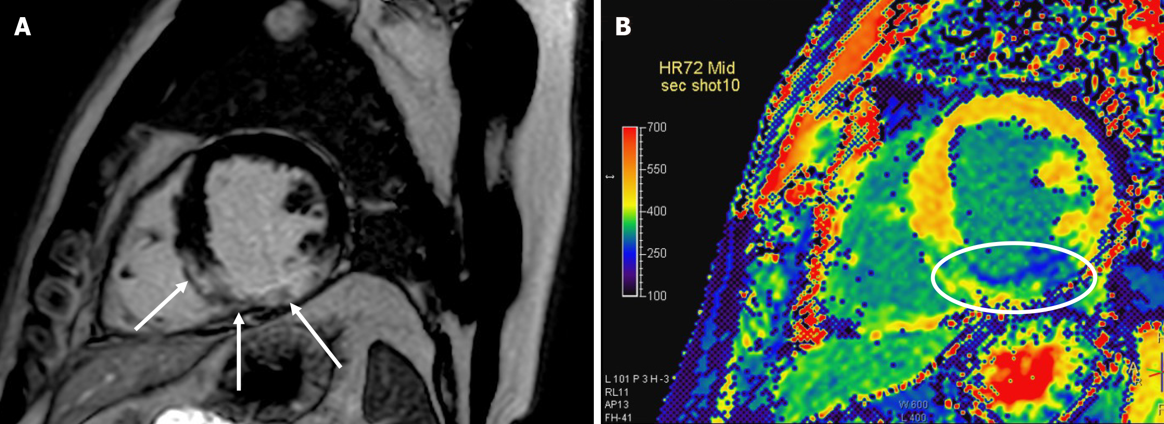
Figure 1 Chronic myocardial infarction presenting with ventricular fibrillation.
A: Heterogeneous late gadolinium enhancement is observed at the midventricular inferior wall (arrows); B: Postcontrast T1 mapping confirms the heterogeneous distribution of gadolinium in the inferior myocardium (circle).
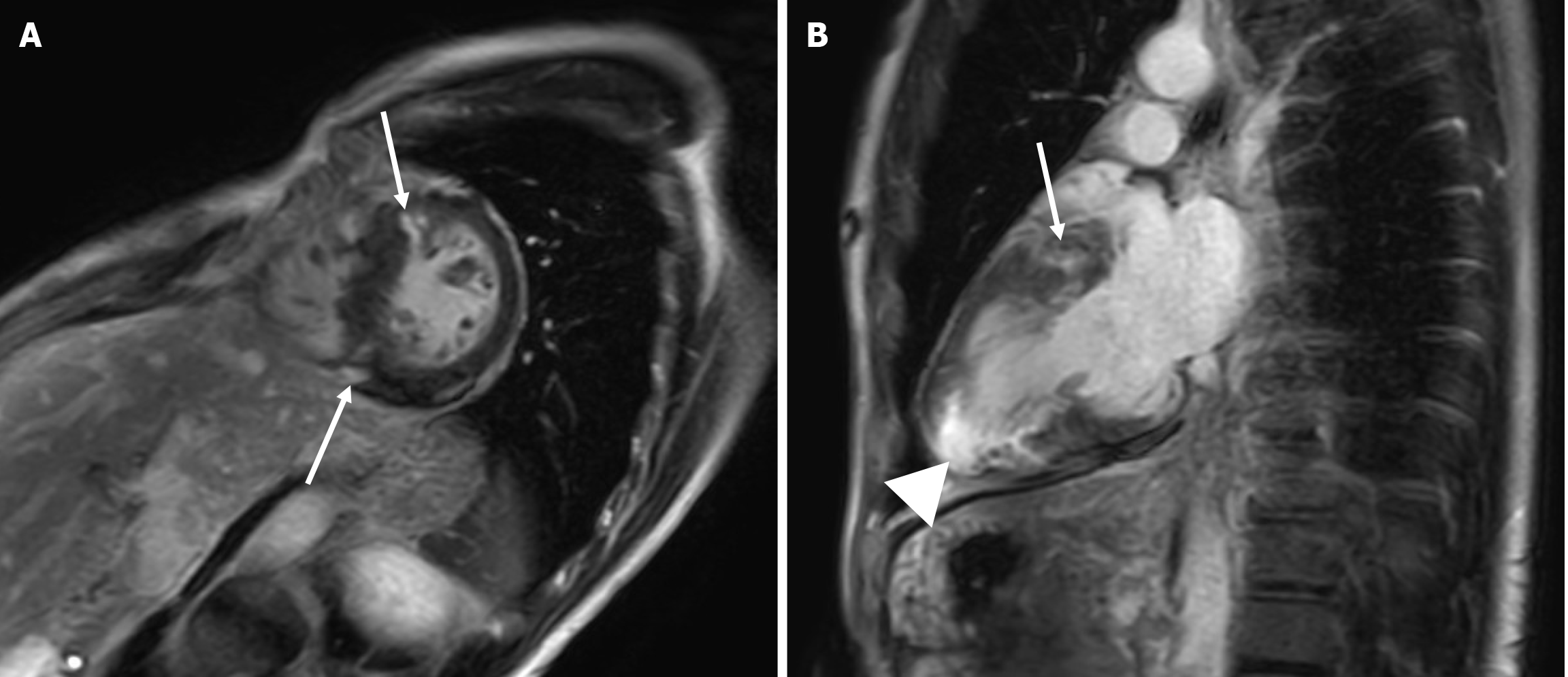
Figure 2 Hypertrophic cardiomyopathy associated with nonsustained ventricular tachycardia.
A: Late gadolinium enhancement (LGE) at the insertion points, a characteristic magnetic resonance imaging feature of hypertrophic cardiomyopathy, is observed (arrows); B: In addition to the typical LGE (arrow), apical aneurysm with LGE is identified, which is related to serious ventricular arrhythmias (arrowhead).
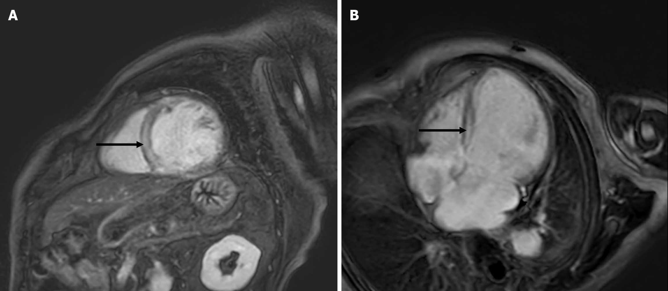
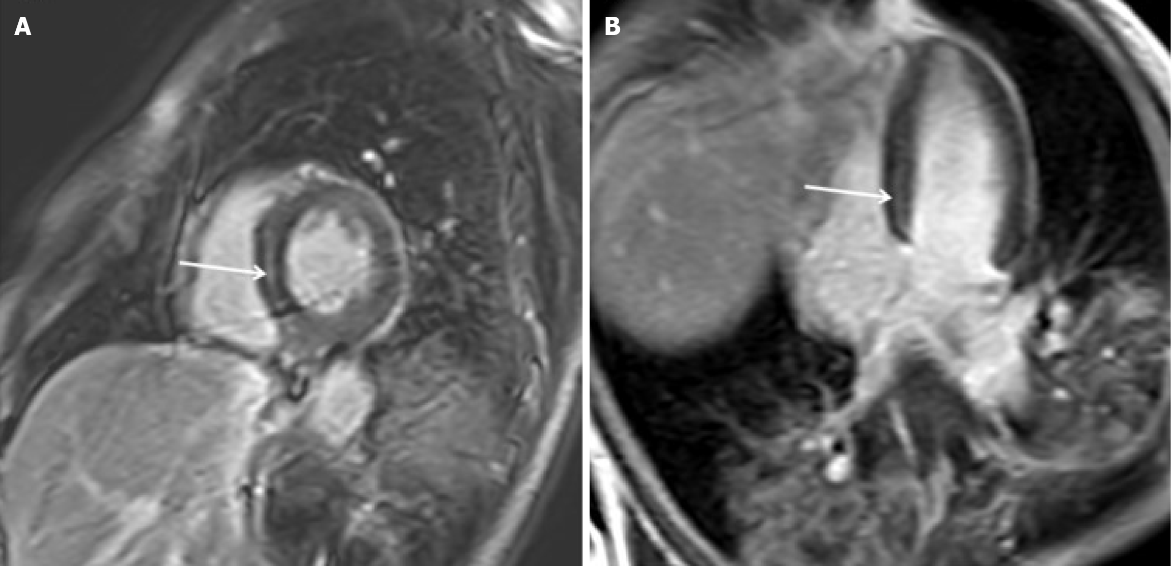
Figure 3 Dilated cardiomyopathy with nonsustained ventricular tachycardia and systolic dysfunction.
A and B: Septal midwall late gadolinium enhancement is observed (arrow). A cardiac resynchronization therapy defibrillator was implanted immediately after magnetic resonance imaging.
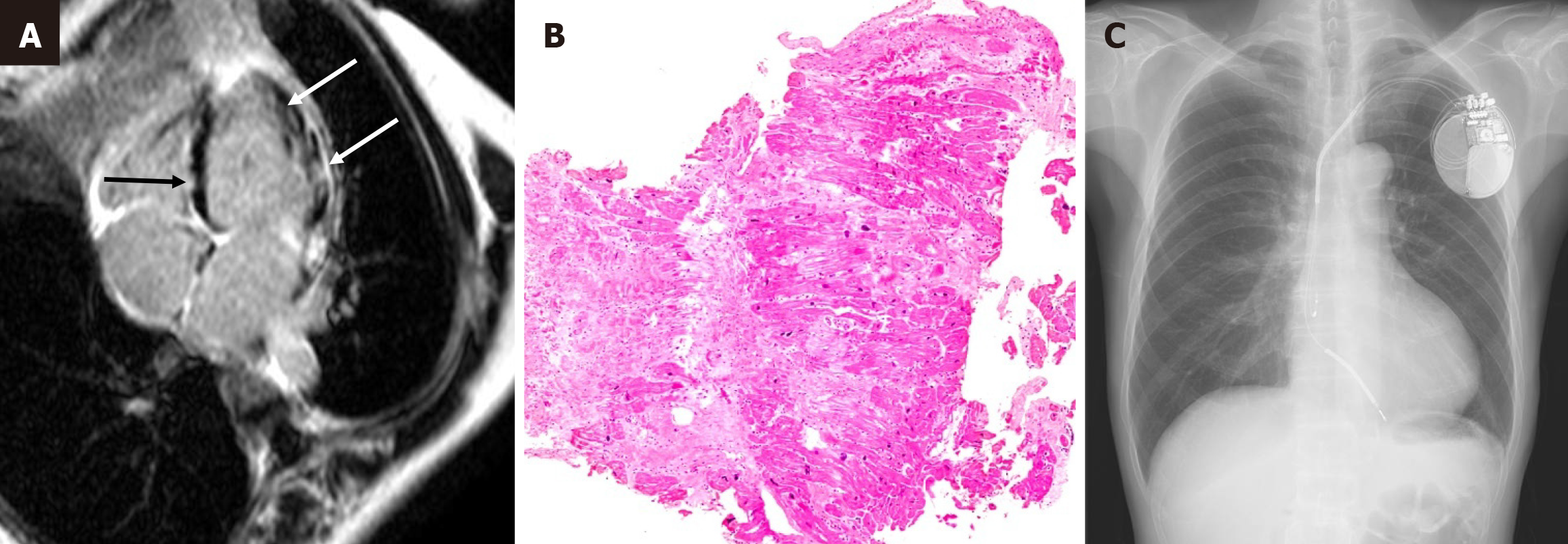
Figure 4 Dilated cardiomyopathy with sustained ventricular tachycardia.
A: Late gadolinium enhancement is observed in both the septal and lateral walls (arrows); B: Endomyocardial biopsy reveals myocardial fibrosis; C: An implantable cardioverter defibrillator was installed.
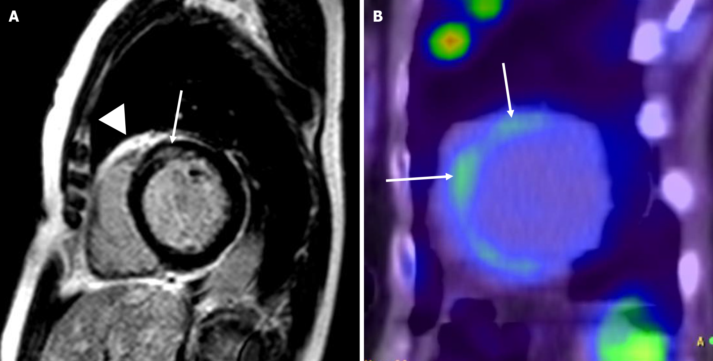
Figure 5 Cardiac sarcoidosis with nonsustained ventricular tachycardia.
A: Late gadolinium enhancement is observed in the anterior wall of both the left (arrow) and right ventricles (arrowhead); B: 18F-fluorodeoxyglucose positron emission tomography shows increased uptake in the anterior and septal wall (arrows).
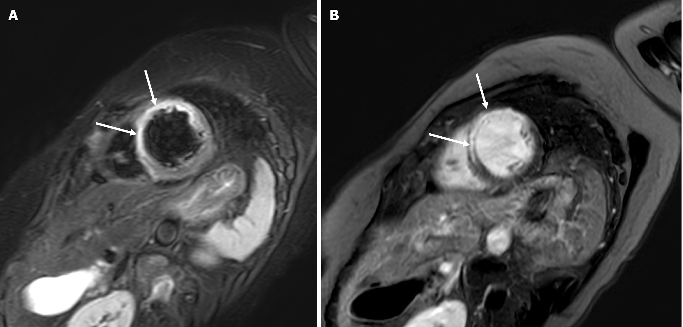
Figure 6 Recurrent acute myocardial infarction presenting with ventricular fibrillation and cardiac arrest.
A: T2-weighted imaging exhibits myocardial edema as high signal intensity (arrows); B: It is difficult to identify acute infarction on late gadolinium enhancement images (arrows).
Figure 7 Coronary vasospasm in a patient with a history of cardiac arrest.
A and B: Septal midwall late gadolinium enhancement is observed on cardiac magnetic resonance imaging before implantable cardioverter defibrillator installation (arrow). Ventricular fibrillation occurred even after implantable cardioverter defibrillator installation in this patient.
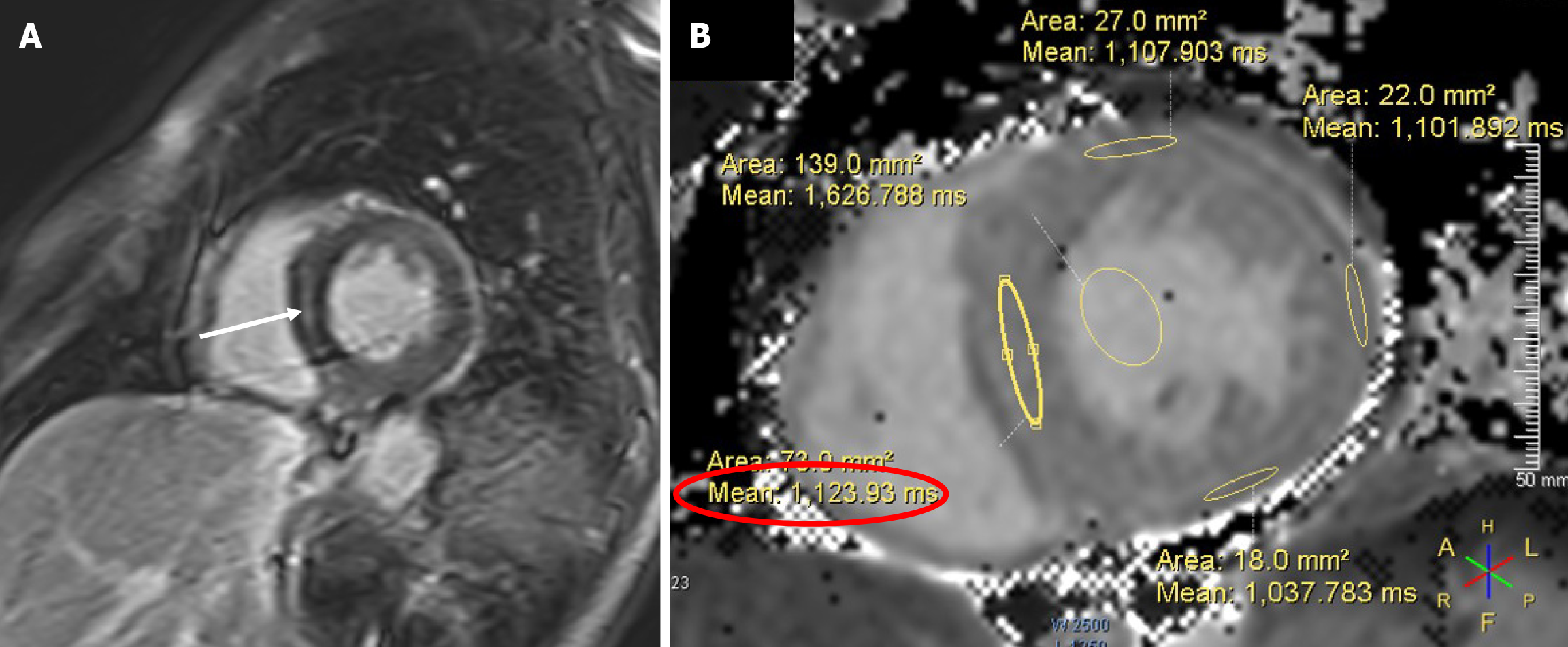
Figure 8 Acute myocarditis associated with cardiac arrest after running.
A: Late gadolinium enhancement is identified in the interventricular septum (arrow); B: T1 mapping reveals elevated T1 values (> 1100 ms, the upper limit of normal in our institution, circle) in the septum, where late gadolinium enhancement is present.
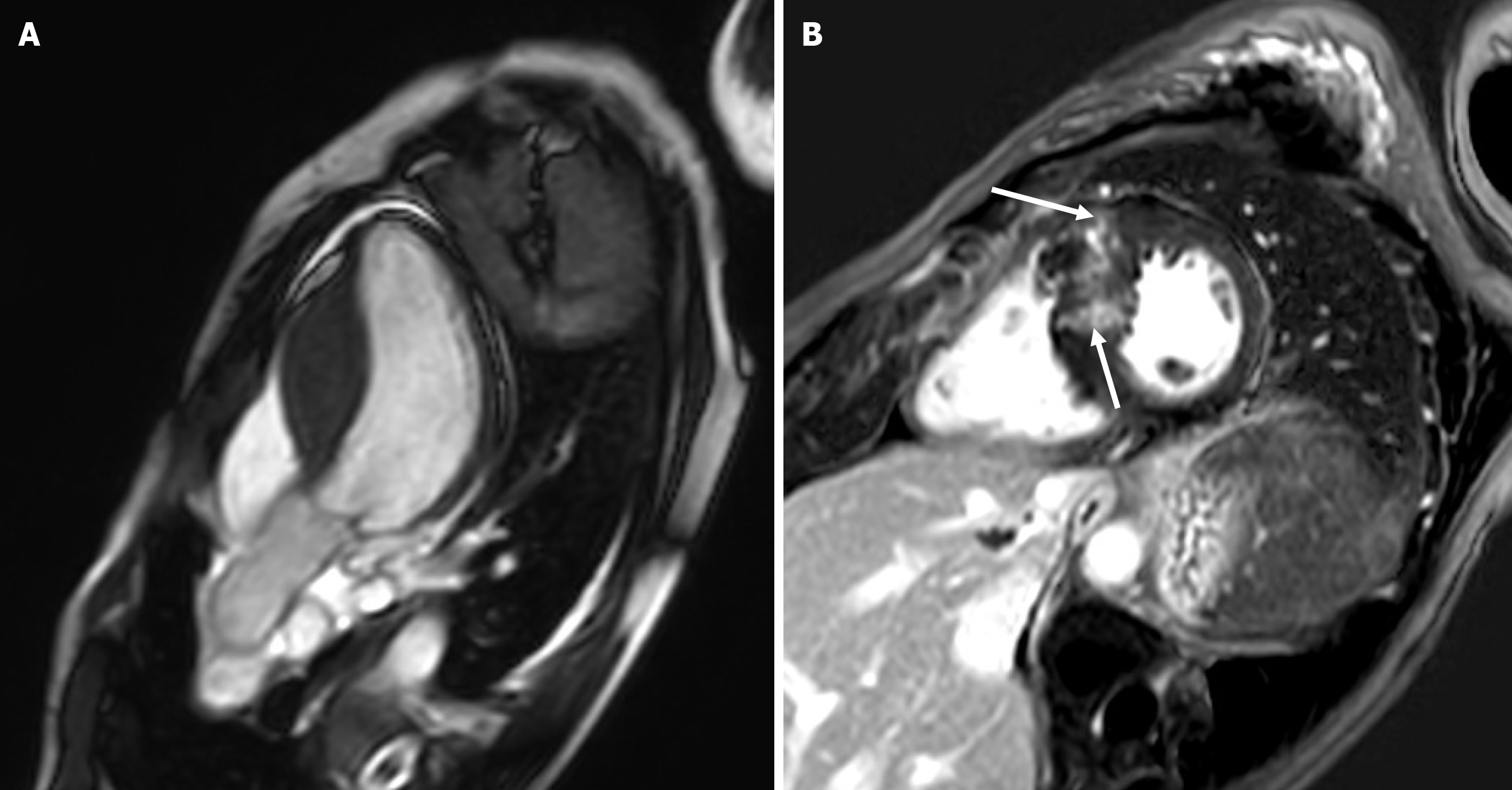
Figure 9 Hypertrophic cardiomyopathy in a patient with out-of-hospital cardiac arrest.
A: Cine magnetic resonance imaging shows a reverse-curve, markedly hypertrophic septum and a thin apex; B: Late gadolinium enhancement is observed in the septal wall (arrows).
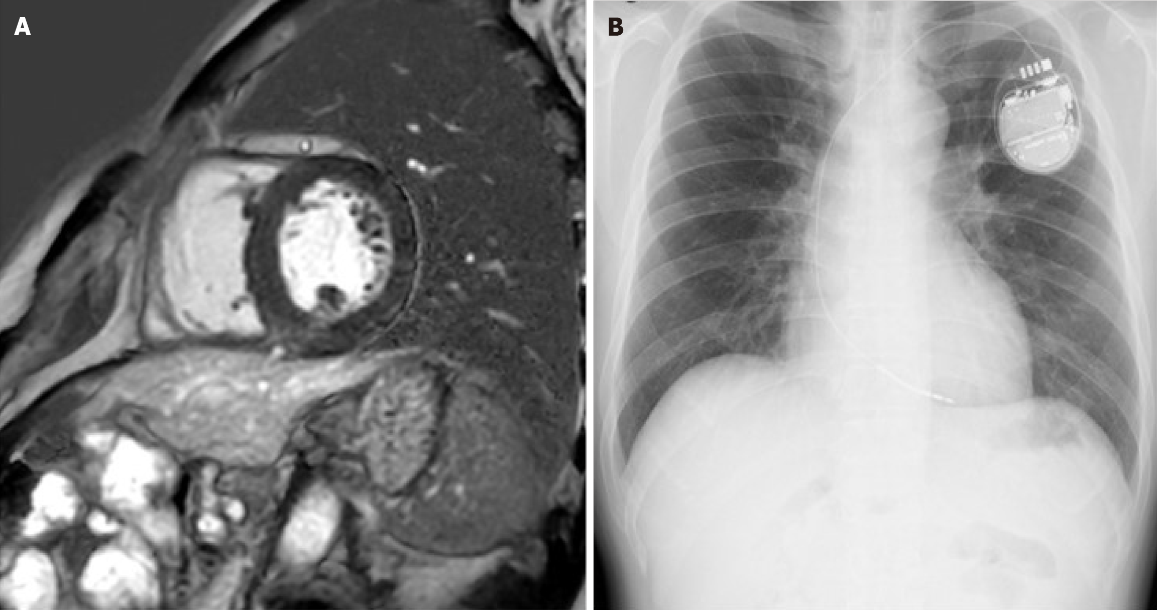
Figure 10 Coronary vasospasm with cardiac arrest during a marathon.
A: No myocardial late gadolinium enhancement is observed; B: An implantable cardioverter defibrillator was implanted.
Figure 11 End-stage hypertrophic cardiomyopathy who died suddenly 6 months after the magnetic resonance imaging study.
A and B: Extensive late gadolinium enhancement is observed at antemortem cardiac magnetic resonance imaging (arrows); C: Endomyocardial biopsy reveals myocytes enlargement, myocardial disarray and collagen fiber infiltration.
- Citation: Amano Y, Iso K, Suzuki Y, Tachi M. Cardiac magnetic resonance imaging contributing to primary prevention and secondary prevention of sudden cardiac death: Contemporary usefulness and limitations. World J Radiol 2025; 17(7): 107140
- URL: https://www.wjgnet.com/1949-8470/full/v17/i7/107140.htm
- DOI: https://dx.doi.org/10.4329/wjr.v17.i7.107140


