©The Author(s) 2025.
World J Radiol. May 28, 2025; 17(5): 107141
Published online May 28, 2025. doi: 10.4329/wjr.v17.i5.107141
Published online May 28, 2025. doi: 10.4329/wjr.v17.i5.107141
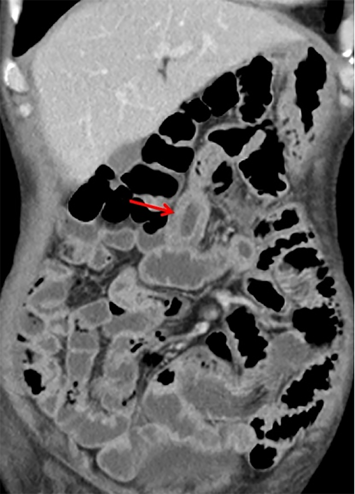
Figure 1 Computed tomography image of the small intestine showing thickening of the jejunal wall and flattening of the intestinal folds.
Figure 2 Histopathological examination of a small intestine mucosal biopsy.
A: Lymphoid tissue hyperplasia in the lamina propria of the small intestinal mucosa on the efferent loop (hematoxylin-eosin staining; original magnification, × 40); B: Lymphocytes are diffusely distributed between the glandular structures, without destroying the glandular architecture (hematoxylin-eosin stain; original magnification, × 400).
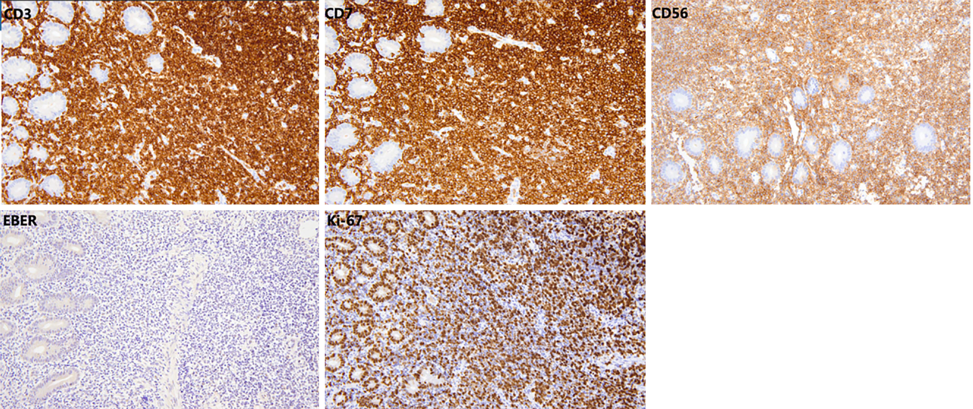
Figure 3 Immunohistochemical analysis showing CD3(++), CD7(++), CD56(++), and Ki-67 (index 70%).
EBER in situ hybridization was negative.
- Citation: Jiang S, Wang LJ, Jia CW, Zhang W, Wang W, Li HL, Sun XH, Qu X, Kang L. Indolent NK-cell lymphoproliferative disorder of the gastrointestinal tract complicated by protein-losing enteropathy: A case report. World J Radiol 2025; 17(5): 107141
- URL: https://www.wjgnet.com/1949-8470/full/v17/i5/107141.htm
- DOI: https://dx.doi.org/10.4329/wjr.v17.i5.107141


