©The Author(s) 2025.
World J Radiol. May 28, 2025; 17(5): 105785
Published online May 28, 2025. doi: 10.4329/wjr.v17.i5.105785
Published online May 28, 2025. doi: 10.4329/wjr.v17.i5.105785
Figure 1 Leathery skin changes.
A and B: Leathery skin changes in the axilla; C: Leathery skin changes in the hip area.
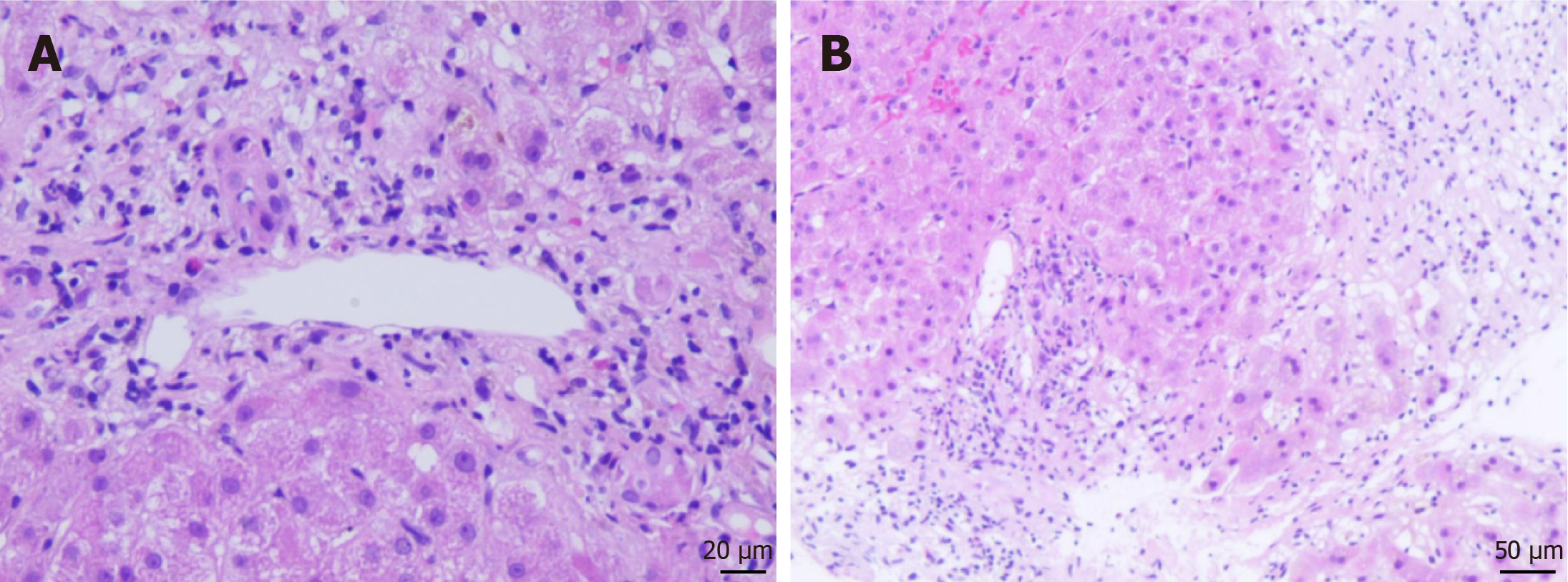
Figure 2 Histological examination of liver biopsy specimen.
A and B: Representative H&E staining of liver biopsy specimen. Scale bar = 20 μm (A), 50 μm (B).
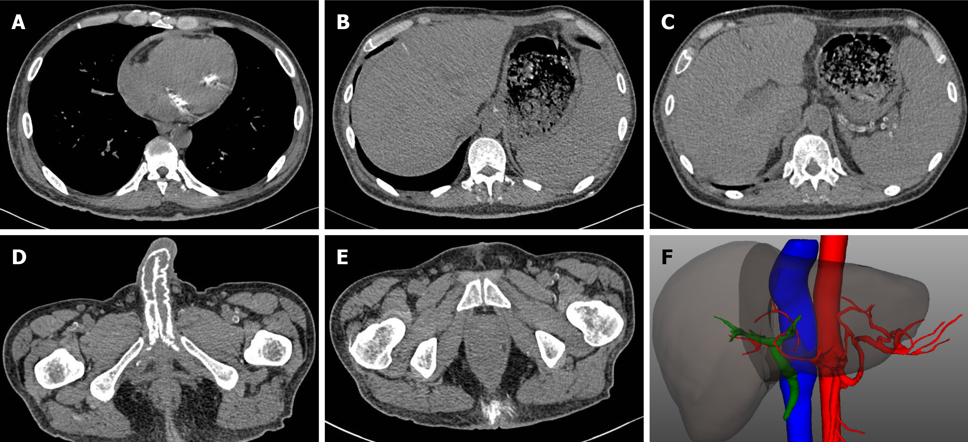
Figure 3 Computed tomography images.
A: Calcification of the bicuspid valve; B: Calcium deposit in the liver; C: Calcification of the splenic artery; D: Calcification in the penis; E: Calcium deposition in the subcutaneous tissue of hip area; F: 3D reconstruction of computed tomography showing occlusion of branches of hepatic arteries.
- Citation: Wei XL, Zhang YW, Han M, Sun CJ, Lai GZ, Tang SG, Ye RJ, Xu HQ, Wu LW, Xia WZ. Calciphylaxis following liver transplantation in a patient with end-stage renal disease: A case report. World J Radiol 2025; 17(5): 105785
- URL: https://www.wjgnet.com/1949-8470/full/v17/i5/105785.htm
- DOI: https://dx.doi.org/10.4329/wjr.v17.i5.105785


