©The Author(s) 2025.
World J Radiol. Jan 28, 2025; 17(1): 101221
Published online Jan 28, 2025. doi: 10.4329/wjr.v17.i1.101221
Published online Jan 28, 2025. doi: 10.4329/wjr.v17.i1.101221
Figure 1
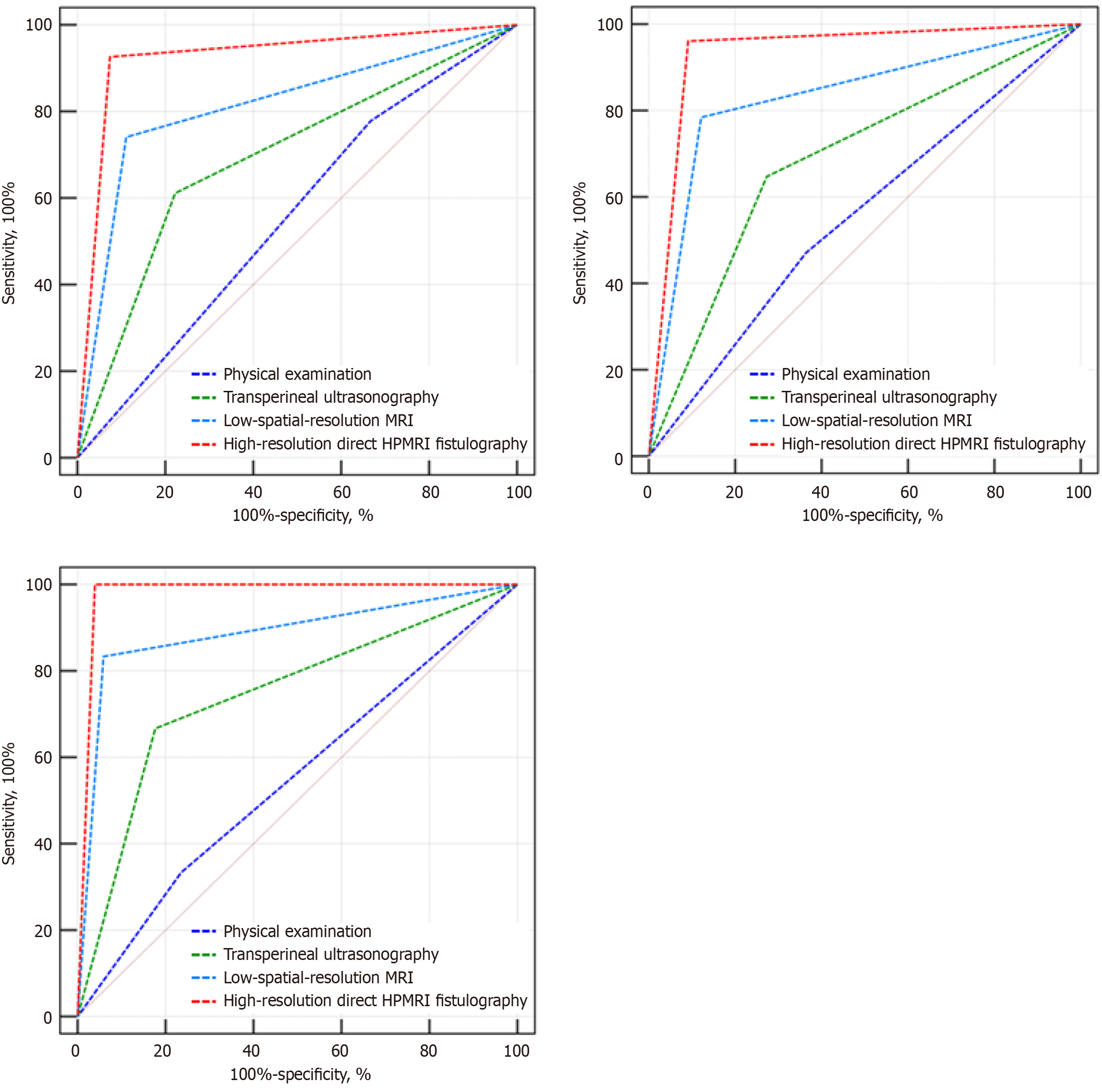
Receiver operating characteristic curves of high-resolution direct hydrogen peroxide-enhanced magnetic resonance imaging fistulography, trans-perineal ultrasonography, low-spatial-resolution magnetic resonance imaging, and physical examination for detecting internal opening, fistula track, and perianal abscess, respectively.
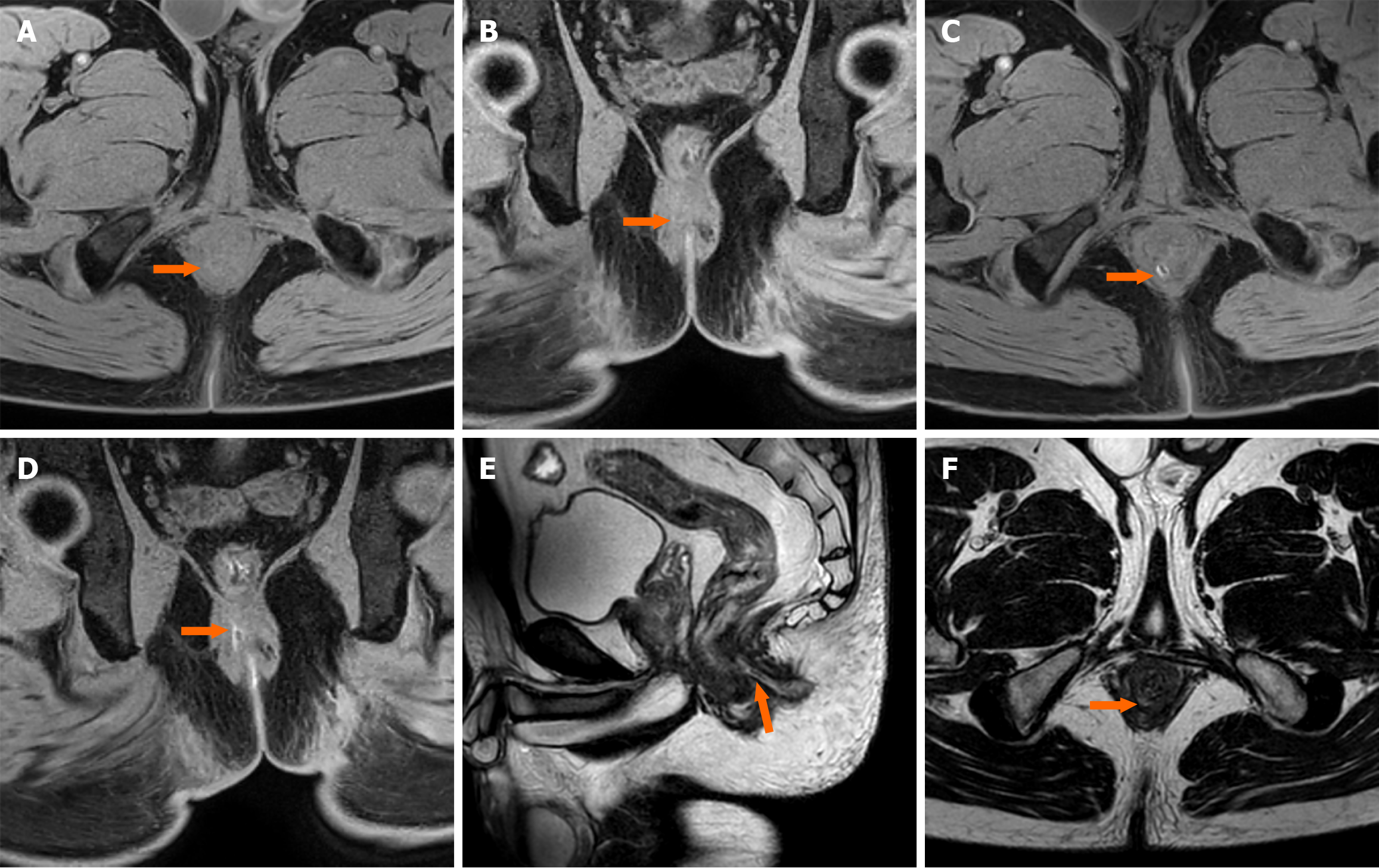
Figure 2 Male patient.
A and B: Oblique axial (A) and oblique coronal (B) T1WI-mDIXON-TFE water image scans of the anal canal, indicating no obvious abnormal signals in the anal canal periphery; C and D: Oblique axial (C) and oblique coronal (D) T1WI-mDIXON-TFE water image high-resolution magnetic resonance imaging (HPMRI) fistulography scans of the anal canal showing a “double-track” high signal (low and high signal in the center and edges, respectively) between the sphincters, and an internal opening at seven o’clock in the lithotomy position (arrowheads); E: Sagittal 3D-T2WI-FSE high-resolution direct HPMRI fistulography, indicating a high intersphincteric fistula track; F: Oblique axial 3D-T2WI-FSE scan of the anal canal showing a small patch of slightly increased signal in the lithotomy position at approximately seven o’clock.
- Citation: Chang CC, Qiao LH, Zhang ZQ, Tian X, Zhang Y, Cheng WW, Wang X, Yang Q. High-resolution direct magnetic resonance imaging fistulography with hydrogen peroxide for diagnosing anorectal fistula: A preliminary retrospective study. World J Radiol 2025; 17(1): 101221
- URL: https://www.wjgnet.com/1949-8470/full/v17/i1/101221.htm
- DOI: https://dx.doi.org/10.4329/wjr.v17.i1.101221


