©The Author(s) 2024.
World J Radiol. Oct 28, 2024; 16(10): 608-615
Published online Oct 28, 2024. doi: 10.4329/wjr.v16.i10.608
Published online Oct 28, 2024. doi: 10.4329/wjr.v16.i10.608
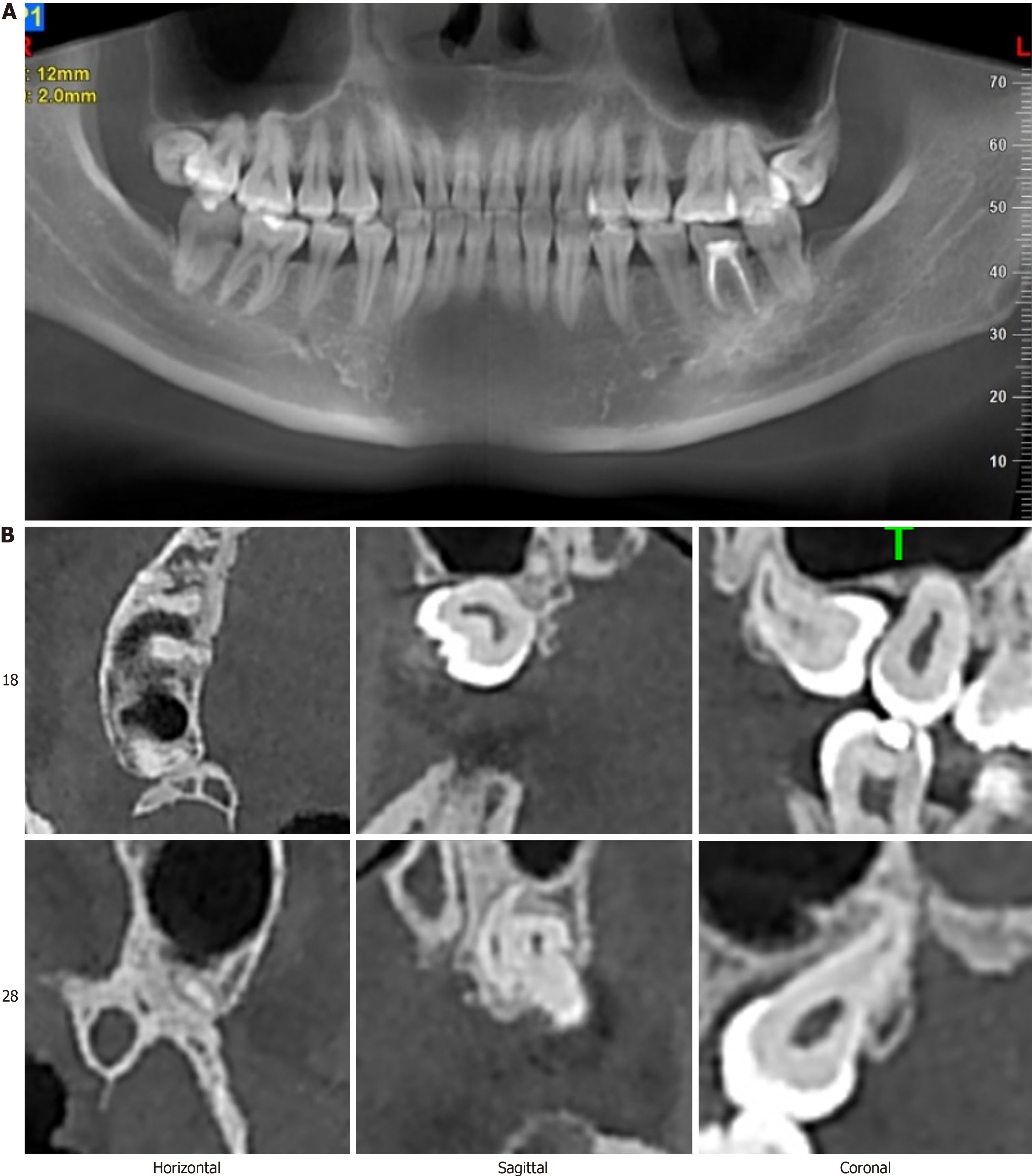
Figure 1 Radiologic data for evaluating the risk of maxillary third molar extraction.
Figure 2 Preoperative imaging.
A: Surface slice; B: Cone-beam computed tomography images at three levels. In the sagittal view, a portion of the crown of tooth 18 is in contact with the maxillary sinus, while in both sagittal and coronal views, part of the root of tooth 28 is seen to abut the maxillary sinus.
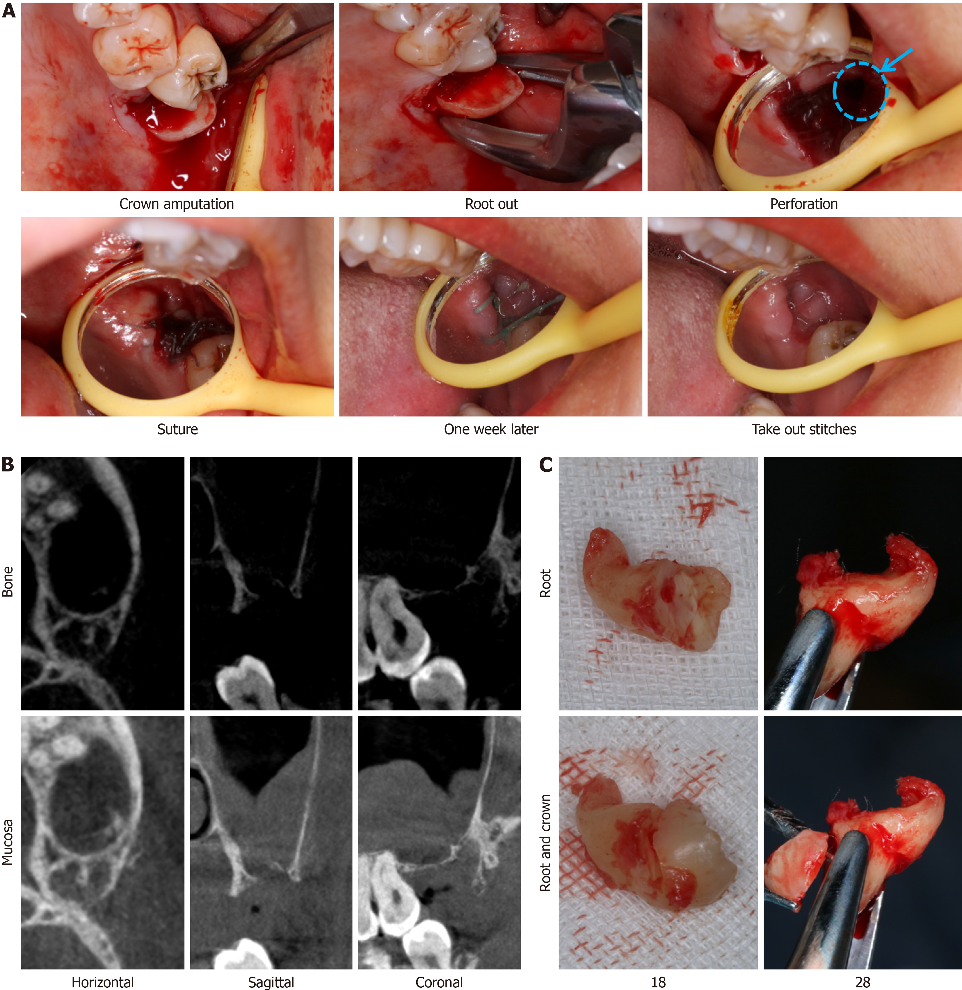
Figure 3 Intraoperative and postoperative imaging.
A: Intraoral image showing a maxillary sinus perforation (blue circle); B: Radiographs obtained two weeks postoperatively; C: Extracted tooth showing the junction of the root and crown.
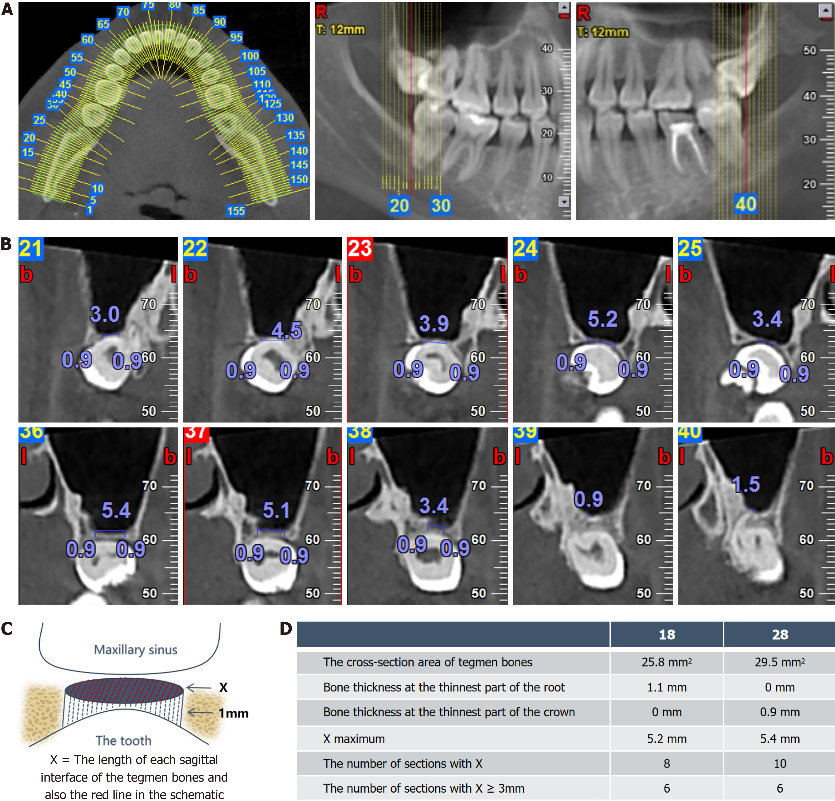
Figure 4 Digital imaging analysis of key factors in cone-beam computed tomography.
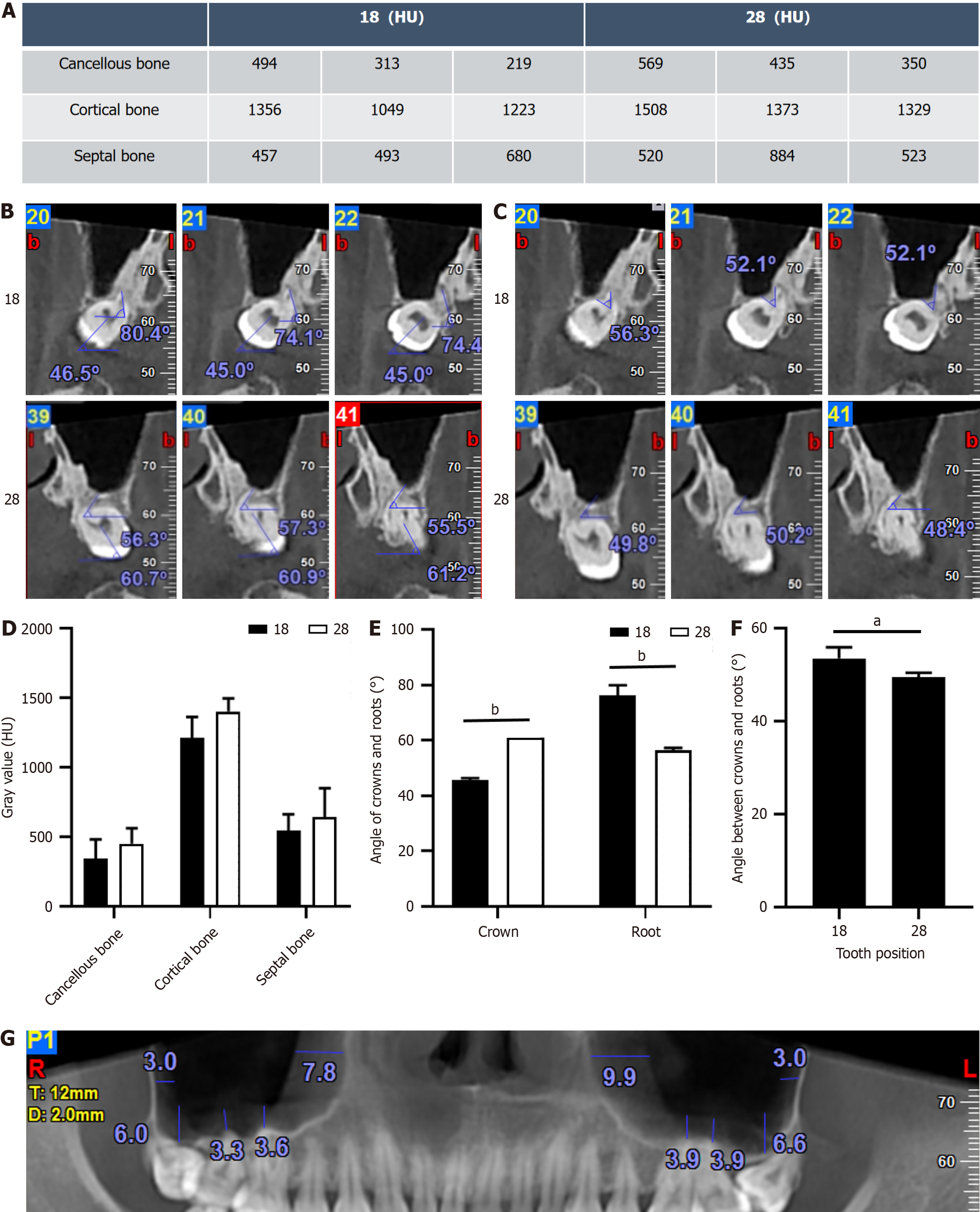
Figure 5 Digital imaging analysis of secondary factors in cone-beam computed tomography.
A: Measurement of gray values; B: Angle measurement between the crown and root relative to the jaw plane; C: Angle measurement between crowns and roots; D: Statistical analysis of gray values at different positions; E: Statistical analysis of the angle between the crown and root relative to the Frankfort plane; F: Statistical analysis of the angle between crowns and roots; G: Measurement of maxillary sinus mucosa. aP < 0.05, bP < 0.001.
- Citation: Liu H, Wang F, Tang YL, Yan X. Asymmetric outcomes in bilateral maxillary impacted tooth extractions: A case report. World J Radiol 2024; 16(10): 608-615
- URL: https://www.wjgnet.com/1949-8470/full/v16/i10/608.htm
- DOI: https://dx.doi.org/10.4329/wjr.v16.i10.608


