©The Author(s) 2022.
World J Radiol. Jul 28, 2022; 14(7): 229-237
Published online Jul 28, 2022. doi: 10.4329/wjr.v14.i7.229
Published online Jul 28, 2022. doi: 10.4329/wjr.v14.i7.229
Figure 1 A 63-year-old patient.
A: Hyperintense lesion on T2 weighted image (WI) in the right breast; B: The lesion is enhanced on post-contrast T1WI; C: The dynamic curve of the lesion is type 2. Pathological diagnosis is invasive ductal carcinoma.
Figure 2 A 37-year-old patient.
A: Hyperintense lesion on T2 weighted image (WI) in the right breast; B: The lesion is enhanced on post-contrast T1WI; C: The dynamic curve of the lesion is type 2. Pathological diagnosis is fibroadenoma.
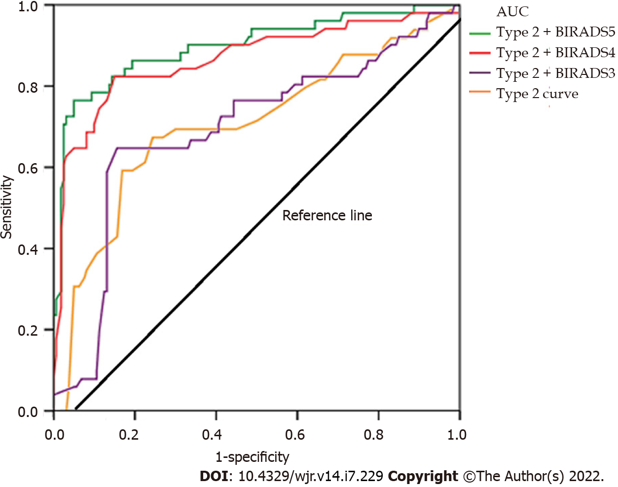
Figure 3 Receiver operating characteristic analysis graph by combining type 2 curves and morphological features for predicting malignancy.
AUC: Area under the curve.
- Citation: Karavas E, Ece B, Aydın S. Type 2 dynamic curves: A diagnostic dilemma. World J Radiol 2022; 14(7): 229-237
- URL: https://www.wjgnet.com/1949-8470/full/v14/i7/229.htm
- DOI: https://dx.doi.org/10.4329/wjr.v14.i7.229


