©The Author(s) 2022.
World J Radiol. Jan 28, 2022; 14(1): 13-18
Published online Jan 28, 2022. doi: 10.4329/wjr.v14.i1.13
Published online Jan 28, 2022. doi: 10.4329/wjr.v14.i1.13
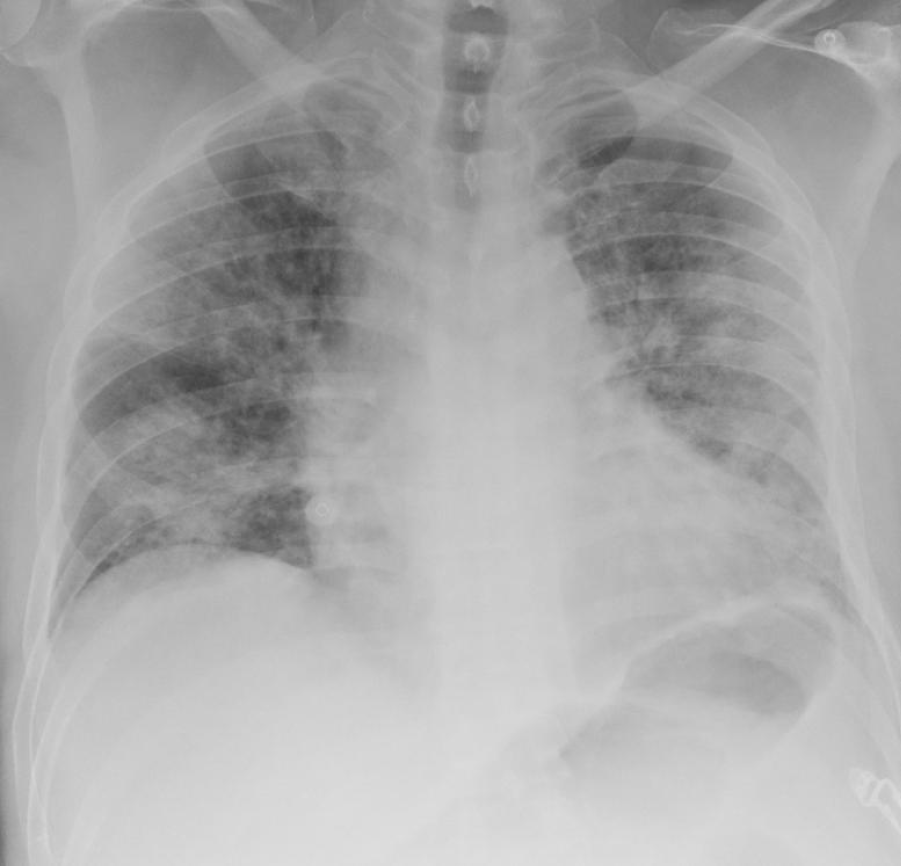
Figure 1 The Chest X-Ray demonstrates multiple bilateral peripheral predominant airspace opacities.
There is no pleural effusion.
Figure 2 Chest X-Ray.
A: Typical appearances of COVID-19 infection: Bilateral peripheral consolidation (1. block arrow), multifocal groundglass opacities (2. straight arrow); B: Some areas of smooth intralobular septal thickening (3. curved arrow).
- Citation: Gangadharan S, Parker S, Ahmed FW. Chest radiological finding of COVID-19 in patients with and without diabetes mellitus: Differences in imaging finding. World J Radiol 2022; 14(1): 13-18
- URL: https://www.wjgnet.com/1949-8470/full/v14/i1/13.htm
- DOI: https://dx.doi.org/10.4329/wjr.v14.i1.13


