©The Author(s) 2020.
World J Radiol. Dec 28, 2020; 12(12): 316-326
Published online Dec 28, 2020. doi: 10.4329/wjr.v12.i12.316
Published online Dec 28, 2020. doi: 10.4329/wjr.v12.i12.316
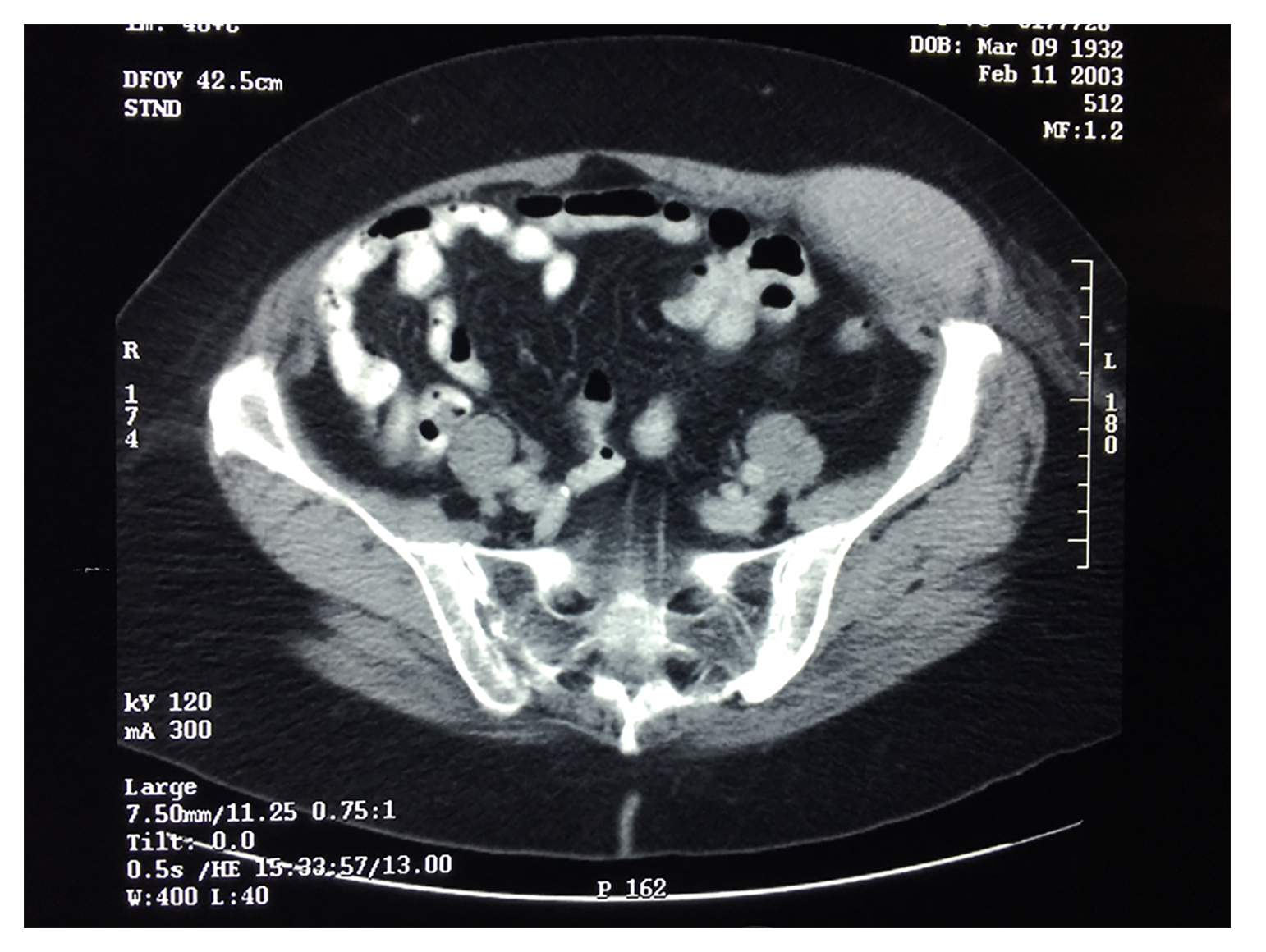
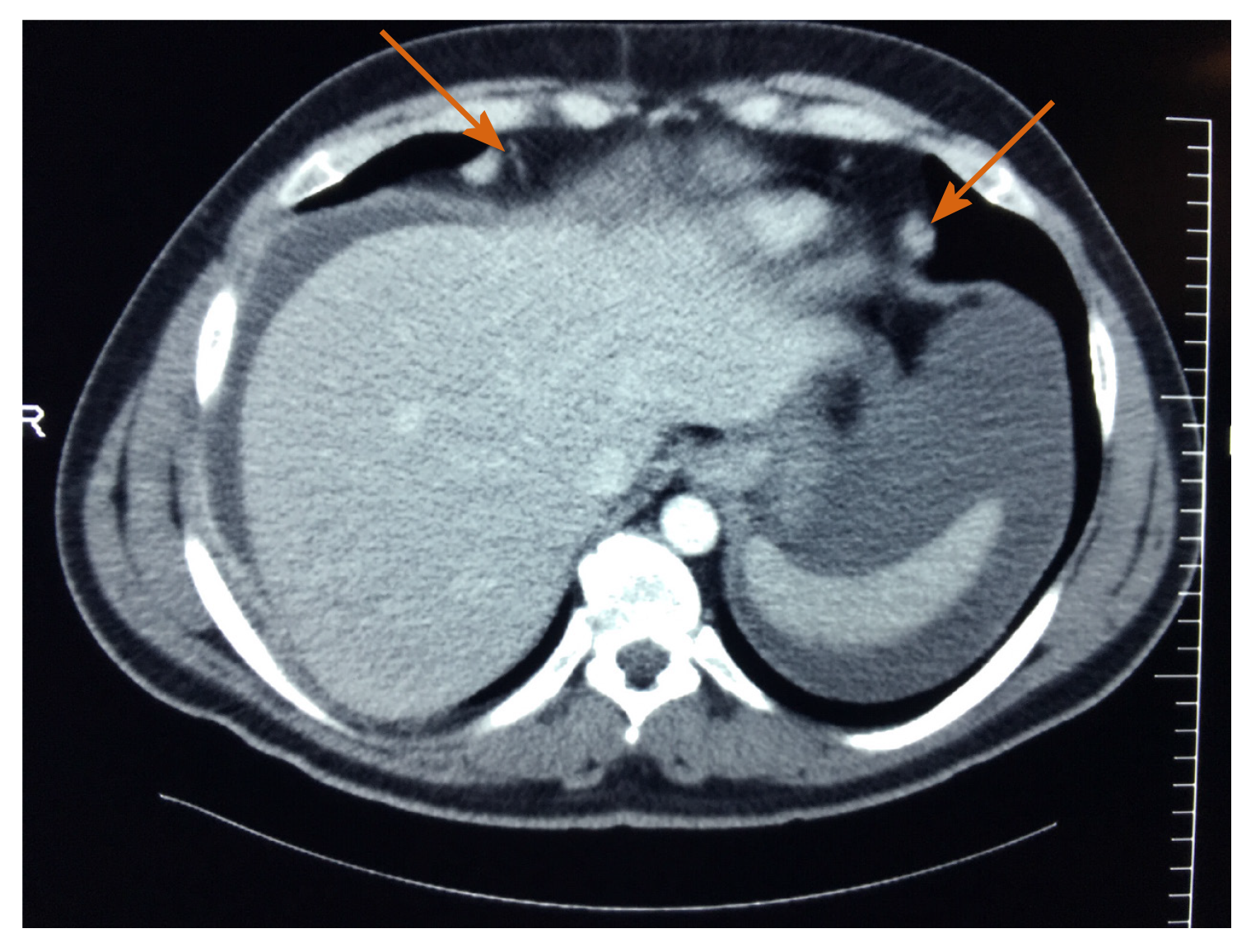
Figure 1 Malignant peritoneal mesothelioma was expanding as a mass within a Spigelian hernia.
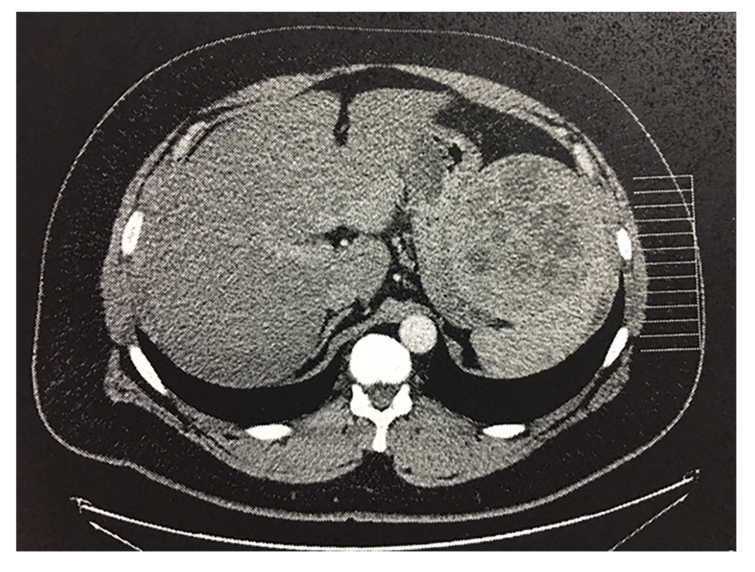
Figure 2 Malignant peritoneal mesothelioma had formed spherical masses contiguous with the spleen.
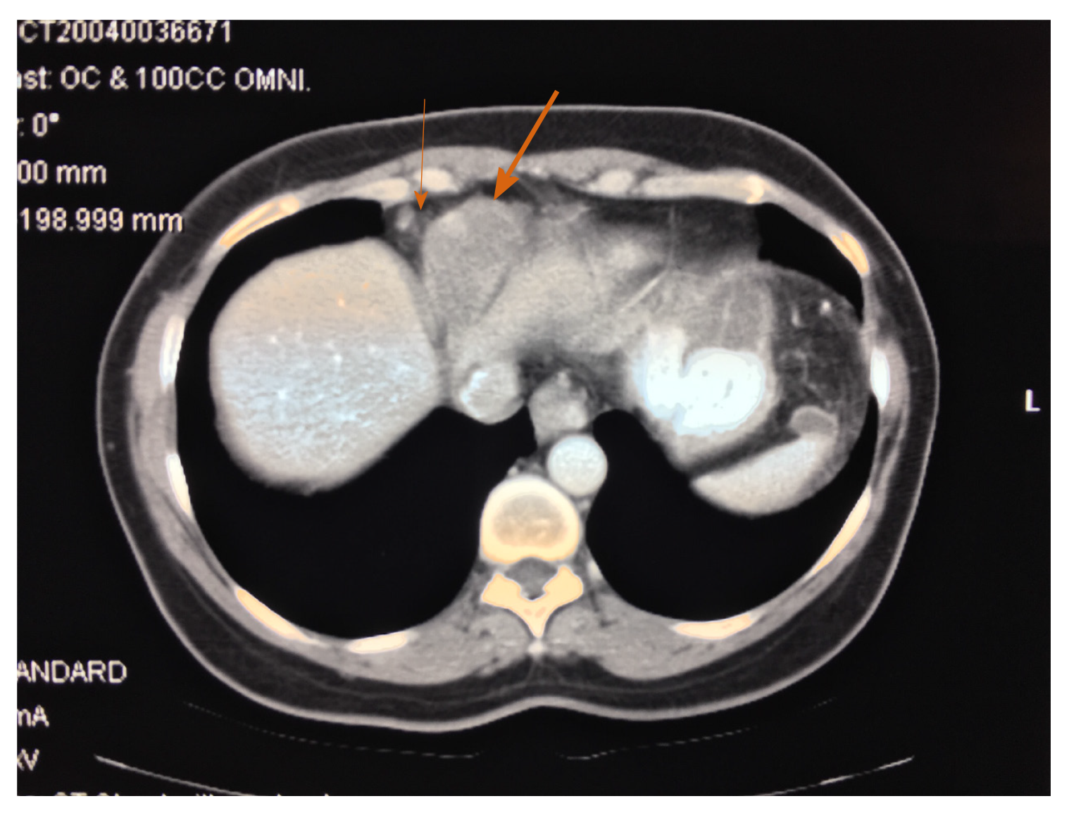
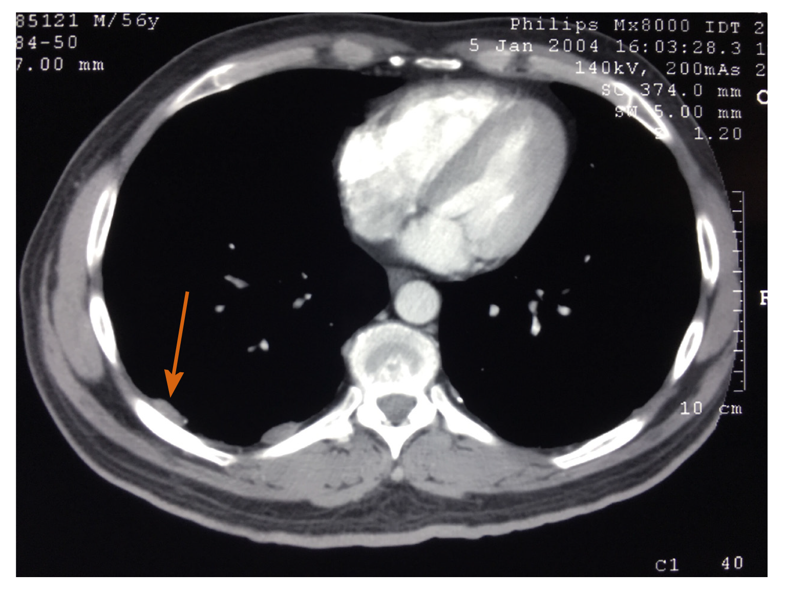
Figure 3 A mass is located directly beneath the diaphragm, invading into the inferior aspect of the pericardial sac and distorting the superior aspect of the liver.
The mass was composed entirely of malignant mesothelioma (large arrow). An enlarged right costophrenic angle lymph node was depicted (small arrow). This disease may be best described as a combined malignant peritoneal and pericardial mesothelioma.
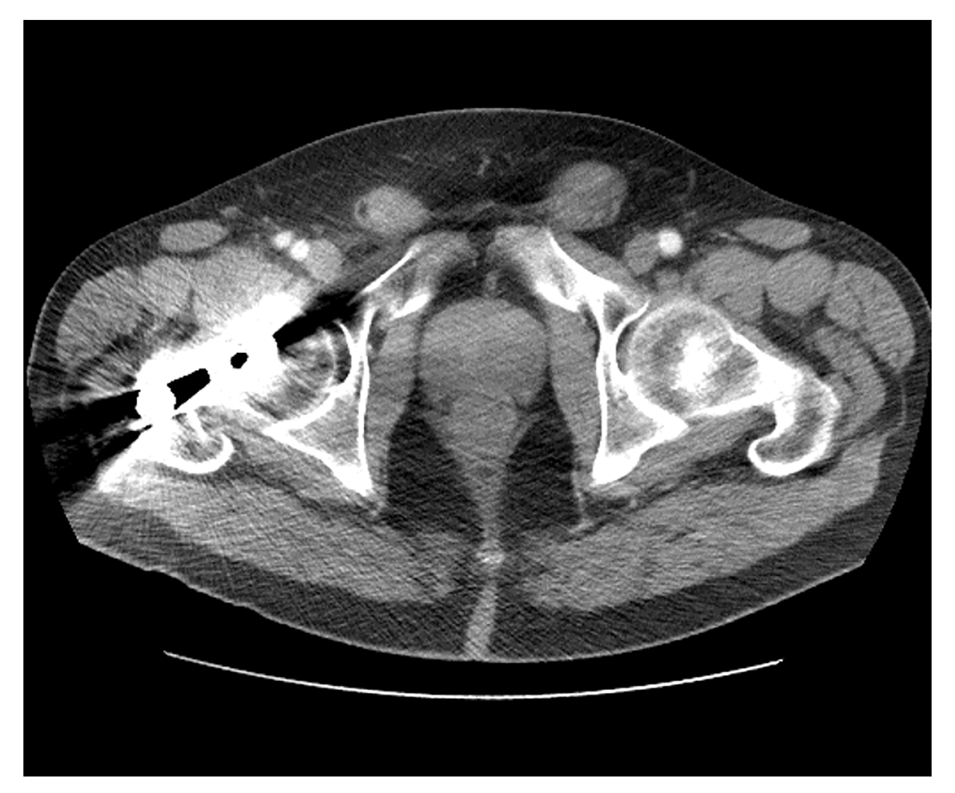
Figure 4 Malignant peritoneal mesothelioma tumor masses were present within the left and right inguinal canal.
The mass on the left was larger and was the patient’s presenting symptom.
Figure 5 In this patient with malignant peritoneal mesothelioma bilateral enlarged cardiophrenic angle lymph nodes were depicted.
Figure 6 In this patient with a confirmed diagnosis of malignant peritoneal mesothelioma, prominent right pleural plaques were present.
He had known direct exposure to asbestos in the past.
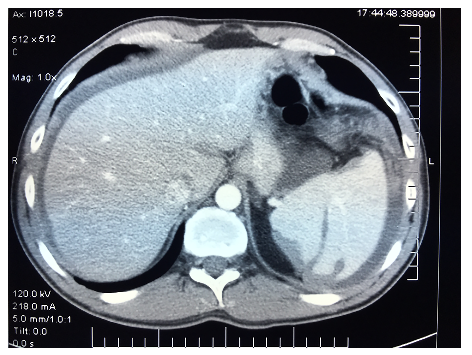
Figure 7 In this patient with a confirmed diagnosis of malignant peritoneal mesothelioma, the spleen was encased by the disease.
Splenic notches are apparent on the surface of the spleen.
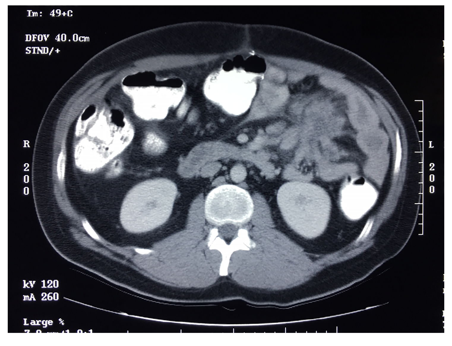
Figure 8 Malignant peritoneal mesothelioma can cause a mass in the left upper quadrant involving the proximal jejunum and mesocolon of the splenic flexure.
- Citation: Sugarbaker PH, Jelinek JS. Unusual radiologic presentations of malignant peritoneal mesothelioma. World J Radiol 2020; 12(12): 316-326
- URL: https://www.wjgnet.com/1949-8470/full/v12/i12/316.htm
- DOI: https://dx.doi.org/10.4329/wjr.v12.i12.316


