Copyright
©The Author(s) 2019.
World J Radiol. May 28, 2019; 11(5): 62-73
Published online May 28, 2019. doi: 10.4329/wjr.v11.i5.62
Published online May 28, 2019. doi: 10.4329/wjr.v11.i5.62
Figure 1 Positron emission tomography/magnetic resonance imaging results by group.
A: Wall thickness across groups; B: Wall area across groups. CVD: Cardiovascular disease.
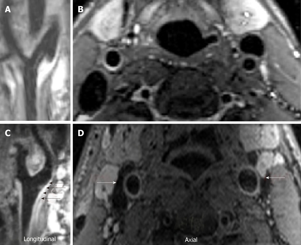
Figure 2 Dark blood magnetic resonance imaging images.
A, B: Healthy vessel in a control subject; C, D: Increased carotid wall thickness (arrows) and area in a cocaine addicted individual. A and C show longitudinal images of the left carotid bifurcation. B and D show axial images of the lateral carotid.
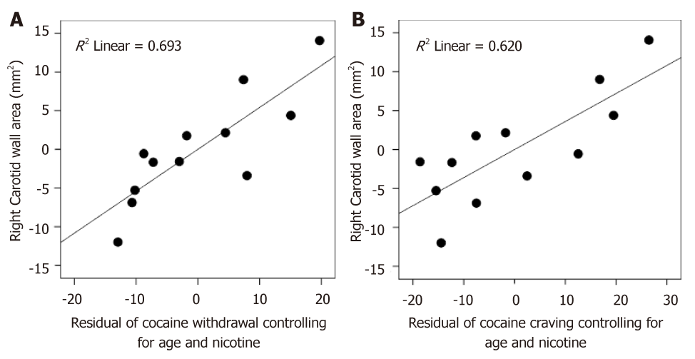
Figure 3 Partial correlation plot.
A: Wall area associations with Cocaine withdrawal symptoms, controlled for age and nicotine; B: Wall area associations with Cocaine craving, controlled for age and nicotine.
- Citation: Bachi K, Mani V, Kaufman AE, Alie N, Goldstein RZ, Fayad ZA, Alia-Klein N. Imaging plaque inflammation in asymptomatic cocaine addicted individuals with simultaneous positron emission tomography/magnetic resonance imaging. World J Radiol 2019; 11(5): 62-73
- URL: https://www.wjgnet.com/1949-8470/full/v11/i5/62.htm
- DOI: https://dx.doi.org/10.4329/wjr.v11.i5.62


