©The Author(s) 2025.
World J Cardiol. Apr 26, 2025; 17(4): 104748
Published online Apr 26, 2025. doi: 10.4330/wjc.v17.i4.104748
Published online Apr 26, 2025. doi: 10.4330/wjc.v17.i4.104748
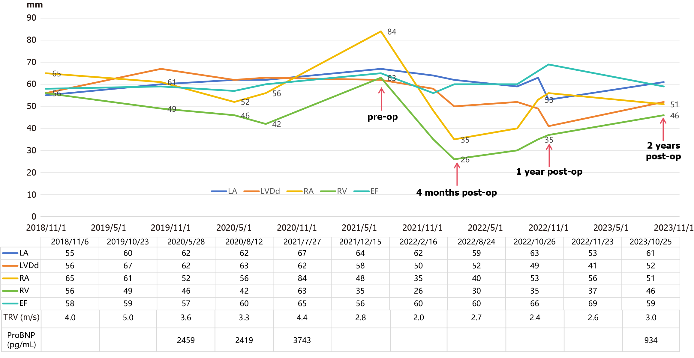
Figure 1 Comparison of preoperative (pre-op) and postoperative (post-op) echocardiographic parameters and pro-BNP levels.
LVDd: Left ventricle end-diastolic dimension; LA: Left atrium; RA: Right atrium; RV: Right ventricle; EF: Ejection fraction; TRV: Tricuspid regurgitant jet velocity.
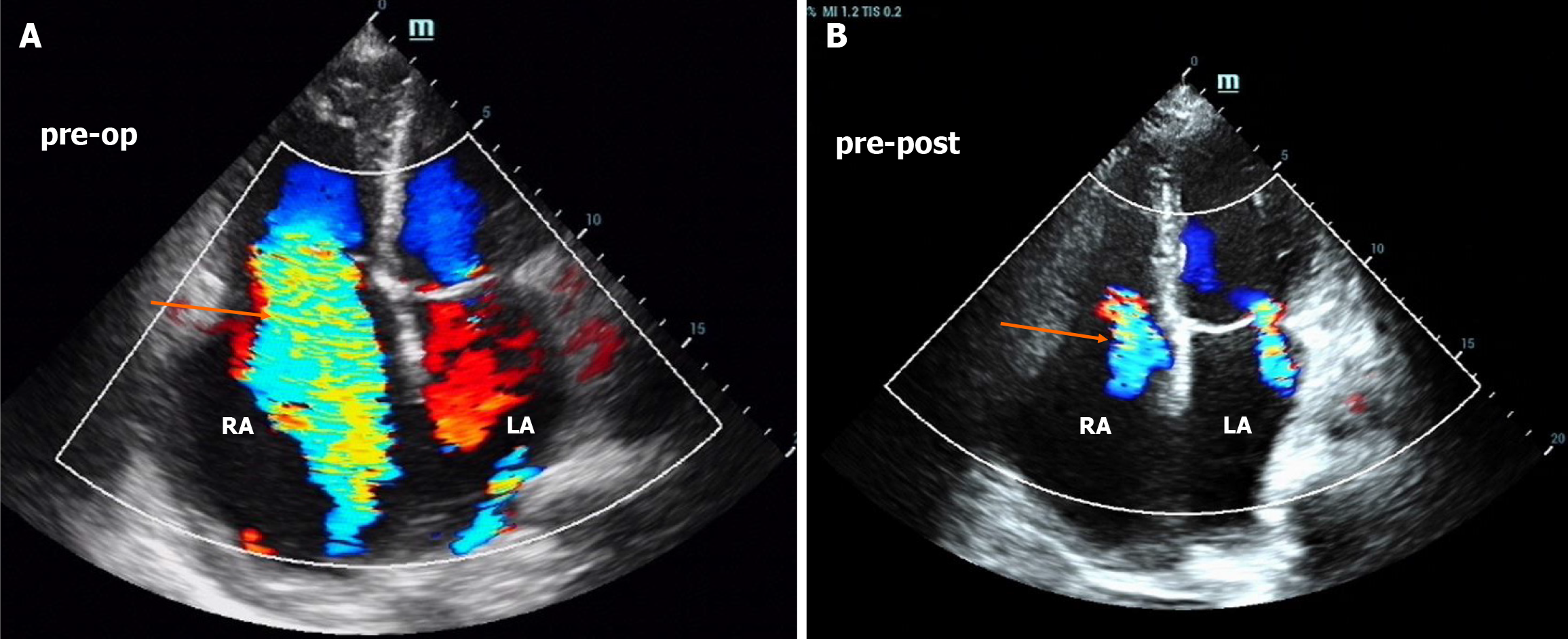
Figure 2 Comparison of preoperative and 4-month postoperative transthoracic Echocardiograms.
A: The diameter of the right atrium (RA) is 84 mm, there is severe tricuspid regurgitation, the maximum flow velocity is 4.4 mL/s, and the estimated pulmonary hypertension is 73 mmHg; B: The diameter of the RA is 35 mm, there is only mild tricuspid regurgitation, and the maximum flow velocity is 2.0 mL/s (the orange arrow represents the tricuspid regurgitation jet).
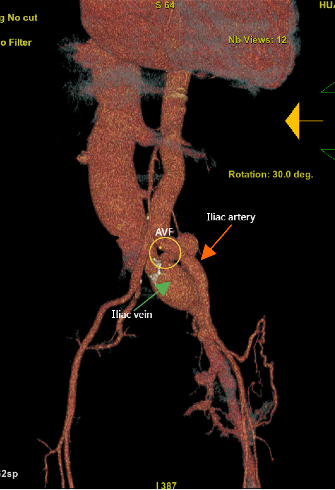
Figure 3
Computed tomography angiography image demonstrating an arteriovenous fistula between the left common iliac artery and vein.
- Citation: He T, He X, Yuan XM. High-output heart failure secondary to iatrogenic arteriovenous fistula: A case report. World J Cardiol 2025; 17(4): 104748
- URL: https://www.wjgnet.com/1949-8462/full/v17/i4/104748.htm
- DOI: https://dx.doi.org/10.4330/wjc.v17.i4.104748


