©The Author(s) 2023.
World J Cardiol. Sep 26, 2023; 15(9): 462-468
Published online Sep 26, 2023. doi: 10.4330/wjc.v15.i9.462
Published online Sep 26, 2023. doi: 10.4330/wjc.v15.i9.462
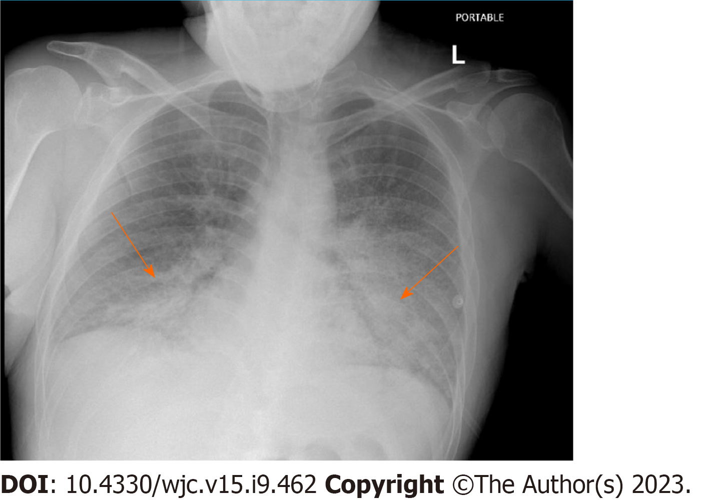
Figure 1 Chest X Ray consistent with acute pulmonary Edema.
Indicating bat wing opacities, interstitial edema, increased cardiothoracic ration and cephalization of pulmonary vessesl.
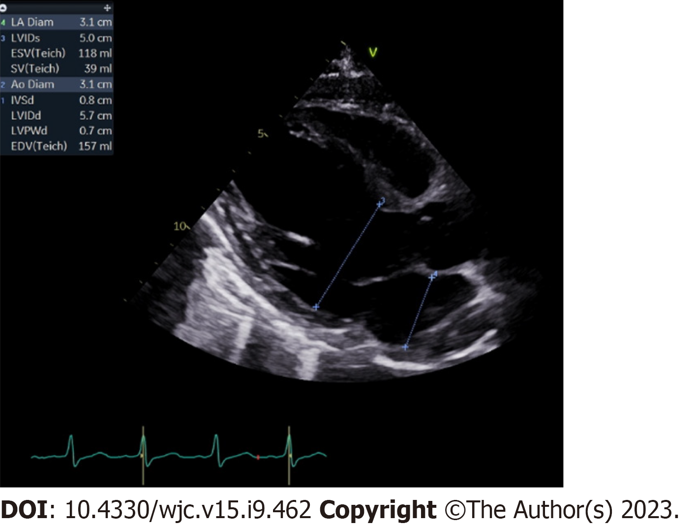
Figure 2 Electrocardiogram indicative of ejection fraction less 20%.
Moderately dilated left ventricle and left atrium.
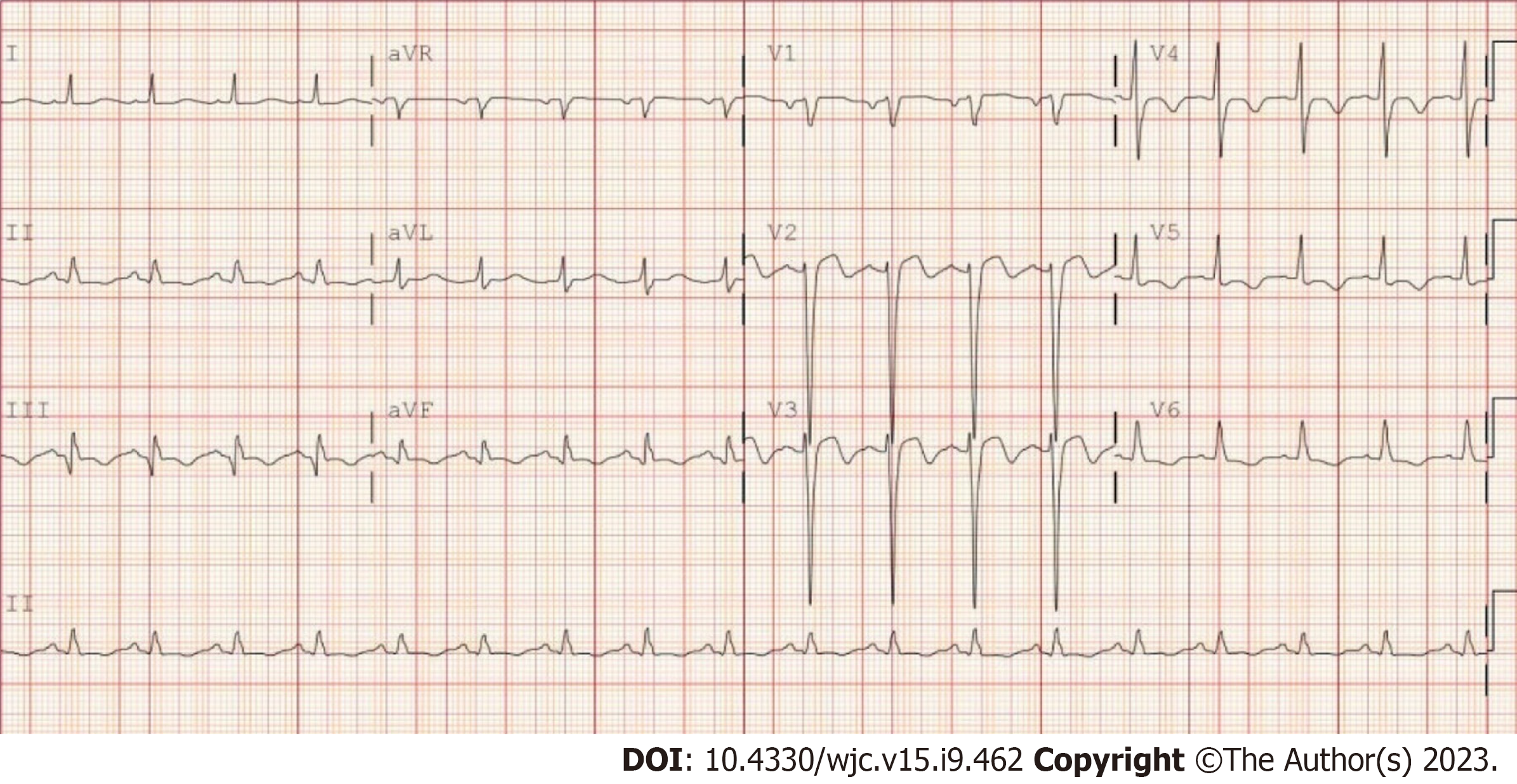
Figure 3 Electrocardiogram showing Biphasic T wave inversion on V2-V3.
T wave inversion in V4-V5.
Figure 4 Coronary angiography report.
A to C: Coronary angiogram report consistent with severe triple vessel disease with stenosis in left anterior descending artery (A), left circumflex artery (B) and right coronary artery (C).
- Citation: Obi MF, Sharma M, Namireddy V, Gargiulo P, Noel C, Hyun C, Gale BD. Variant of Wellen’s syndrome in type 1 diabetic patient: A case report. World J Cardiol 2023; 15(9): 462-468
- URL: https://www.wjgnet.com/1949-8462/full/v15/i9/462.htm
- DOI: https://dx.doi.org/10.4330/wjc.v15.i9.462


