Copyright
©The Author(s) 2023.
World J Cardiol. Oct 26, 2023; 15(10): 479-486
Published online Oct 26, 2023. doi: 10.4330/wjc.v15.i10.479
Published online Oct 26, 2023. doi: 10.4330/wjc.v15.i10.479
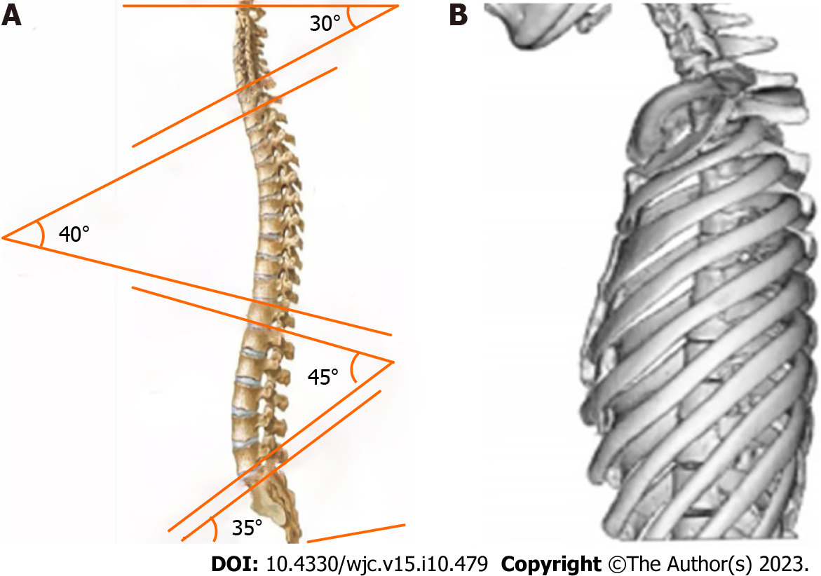
Figure 1 Differentiating between normal individuals and individuals with straight back syndrome with physiological distortion.
A: The human spine typically exhibits physiological curvature; B: In individuals diagnosed with straight back syndrome, this curvature is noticeably absent.
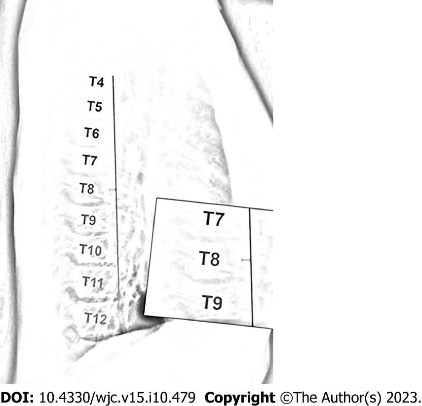
Figure 2 Diagnostic criterion for straight back syndrome on lateral chest radiographs, where the distance from the midpoint of the T8 vertebral body to a vertical line connecting the anterior borders of T4 and T12 is less than 1.
2 cm.
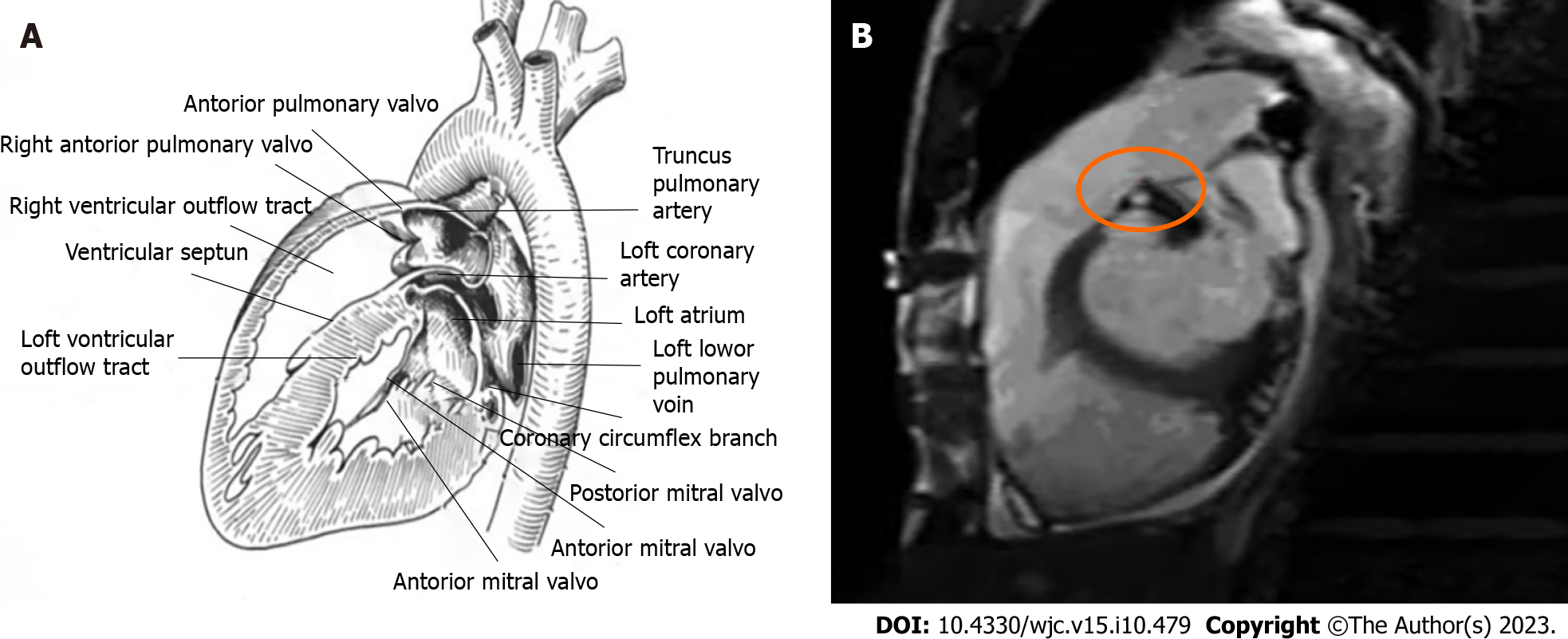
Figure 3 Comparison of right ventricular outflow tract between normal patients and patients with straight back syndrome.
A: Right ventricular outflow tract patency in a normal individual; B: Right ventricular hypertrophy and narrowing of the right ventricular outflow tract in a patient with straight back syndrome.
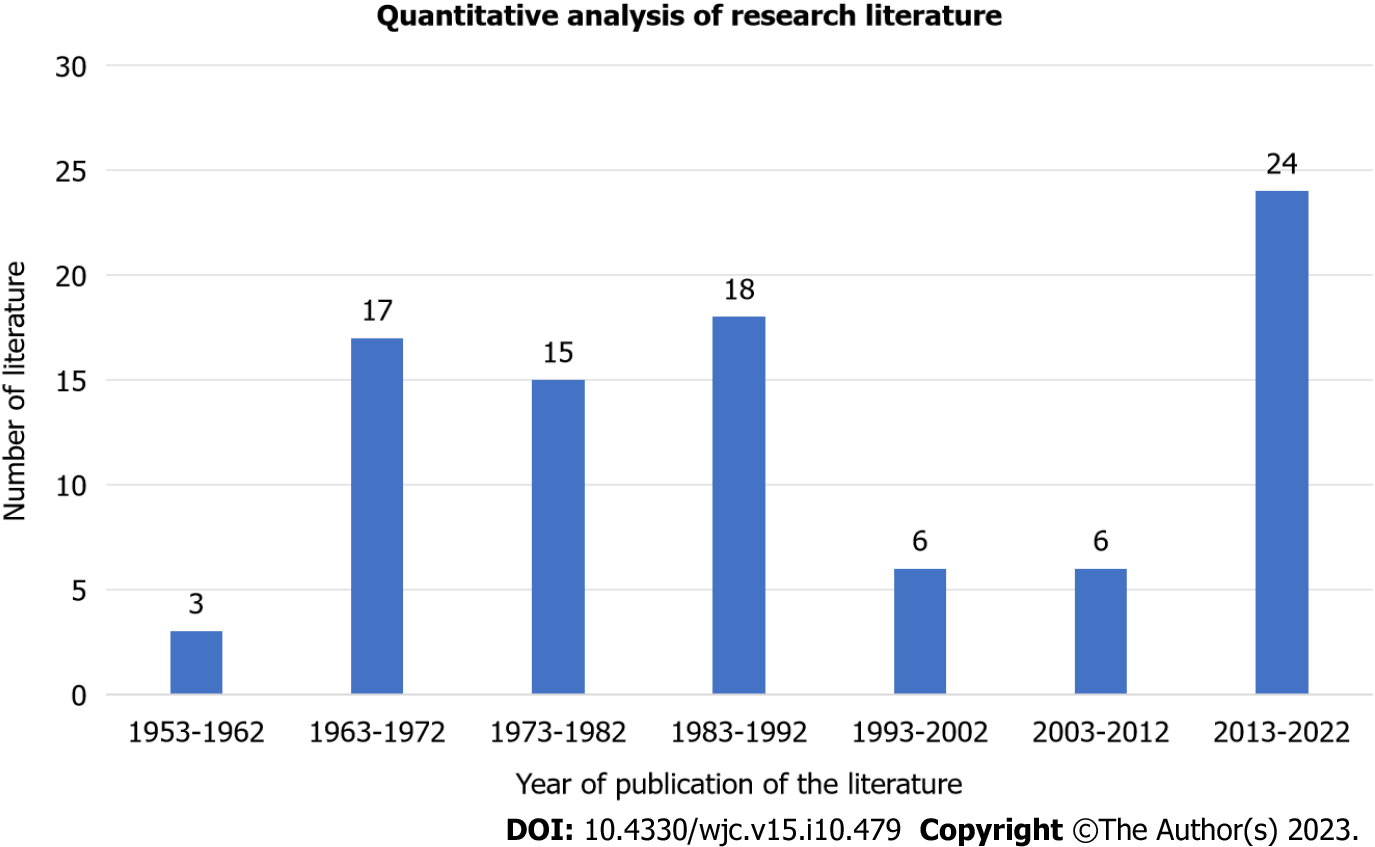
Figure 4 Distribution of straight back syndrome publications from 1953 to 2022.
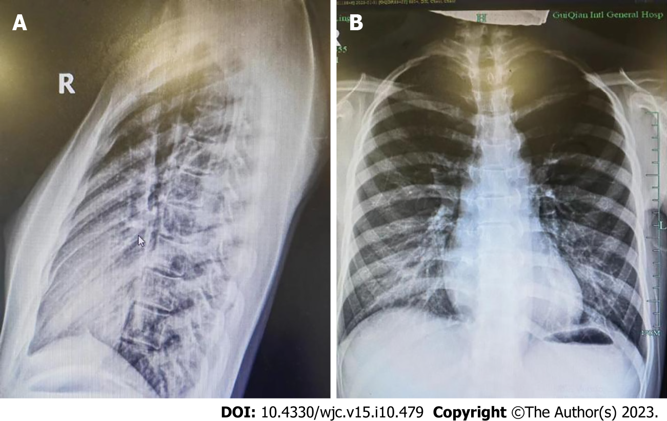
Figure 5 Chest X-ray film of a patient with straight back syndrome.
A: Lateral chest X-ray; B: Anteroposterior chest X-ray.
- Citation: Kong MW, Pei ZY, Zhang X, Du QJ, Tang Q, Li J, He GX. Related mechanisms and research progress in straight back syndrome. World J Cardiol 2023; 15(10): 479-486
- URL: https://www.wjgnet.com/1949-8462/full/v15/i10/479.htm
- DOI: https://dx.doi.org/10.4330/wjc.v15.i10.479


