©2014 Baishideng Publishing Group Inc.
World J Biol Chem. May 26, 2014; 5(2): 141-160
Published online May 26, 2014. doi: 10.4331/wjbc.v5.i2.141
Published online May 26, 2014. doi: 10.4331/wjbc.v5.i2.141
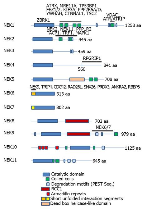
Figure 1 Representation of the domain organization of the eleven human Neks depicting the domain regions for selected protein interactions.
The gene symbols corresponding to interacting proteins are shown above the Neks primary structure regions with which they have been found to interact. The list of interactors is not intended to be complete but is necessarily shorter than the list of all proteins known in the literature to interact with Neks (e.g., see Figure 2), since, for the majority of interactors, the location of interaction in the Neks has not been reported. Different repeated domains have been indicated by the color code at the bottom of the figure. The lengths of the full proteins are indicated by number of amino acids (aa) at the C-terminal of the proteins. At least two isoforms of Nek1, 2, 3 and three of Nek4 and 11, all generated by alternative splicing, have been reported and known functional distinctions have been briefly discussed in the text, where feasible. References for the proteins and their mapped interactors: Nek1[2,13,25]; Nek2[116,121-124]; Nek4[53]; Nek6[3]; Nek9[66]. Nek: Never in mitosis-gene A-related kinases.
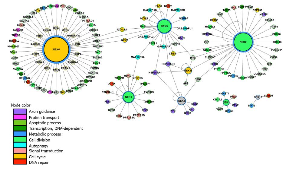
Figure 2 Global interactome of Nek1-11, involving their published interactors.
The proteins color code refers to their main biological function given by the top enriched Gene Ontology[125] biological processes (P≤ 0.05). Common interactors establish crosslinks between Neks, thereby emphasizing their common functional contexts. The protein sizes are depicted proportional to their connectivity degree. The protein-protein interaction network was built for the first neighbors of Neks using the Integrated Interactome System (IIS) platform, developed at National Laboratory of Biosciences, Brazil (http://www.lge.ibi.unicamp.br/lnbio/IIS/) and visualized using the Cytoscape software[126]. Nek: Never in mitosis-gene A-related kinases.
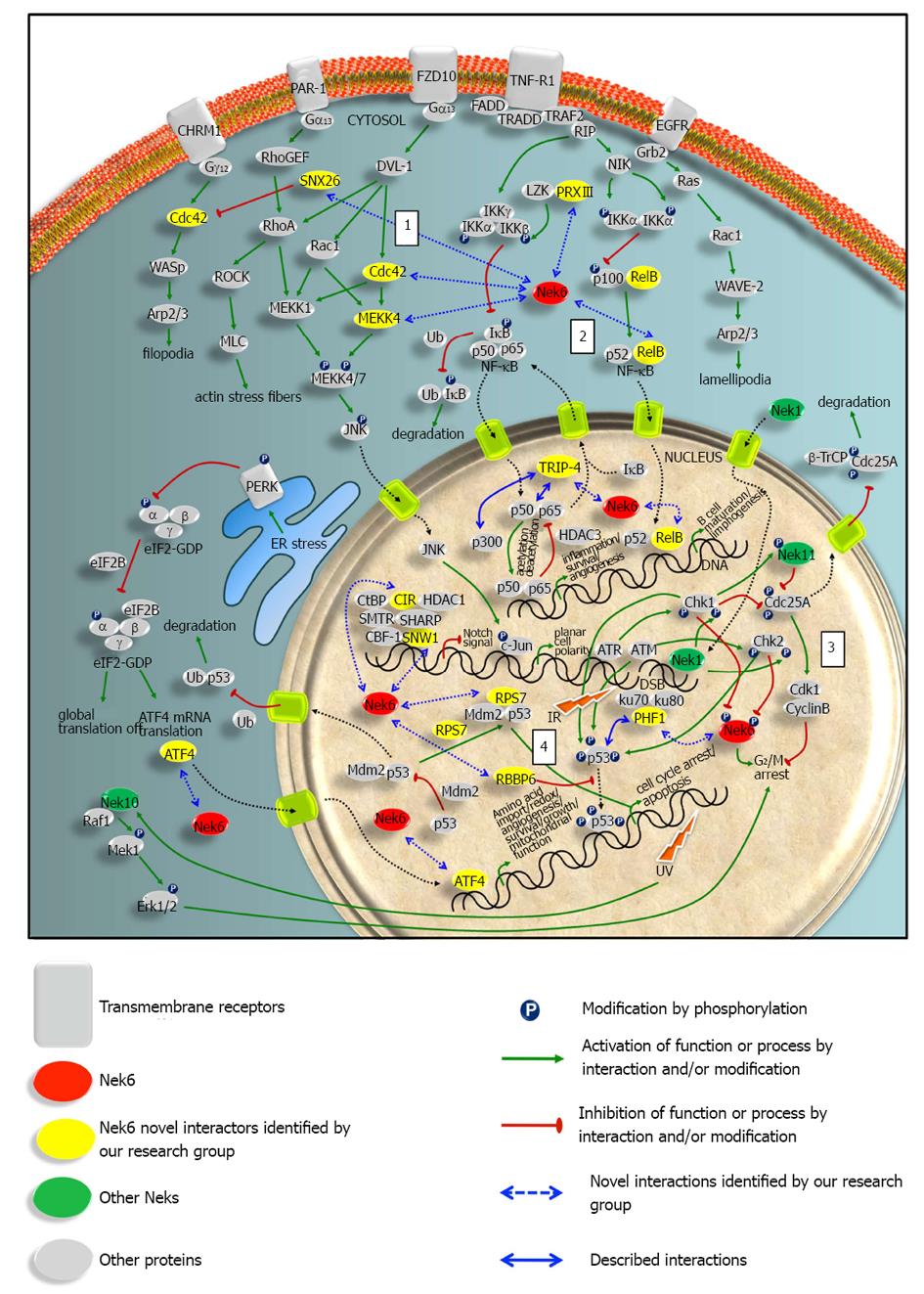
Figure 3 Nek6 interactome and the cellular functional contexts based on its interacting proteins.
The four major pathways discussed in the text are: (1) actin cytoskeleton organization; (2) nuclear factor-κB signaling; (3) DNA damage response; (4) p53 signaling (according to Meirelles et al). See detailed legend for symbols at the bottom of the figure. IR: Ionizing radiation.
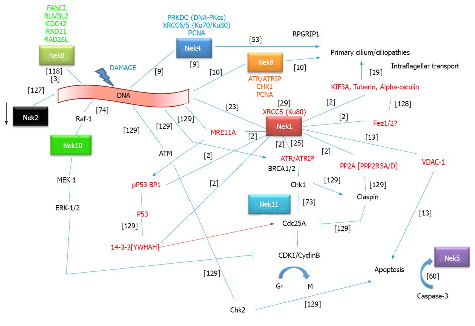
Figure 4 Nek1 interactome and crosstalk with other Neks and protein interactors in the context of the DNA damage response pathways.
Interactions between proteins are depicted as simple lines, activation is depicted as an arrow and inhibition as an arrow with a line as arrowhead. A red arrow for 14-3-3 means that it causes activation by the transport of CDC25 to the nucleus. Nek1 interacted with a specific 14-3-3 isoform called YWHAH[2] (gene symbols inside brackets correspond to the isoforms of those proteins which were described to interact with Nek1). Not necessarily the same specific 14-3-3 protein promotes the indicated functions. Rather, a family characteristic is intended to be assigned. Nek2 kinase activity is inhibited after DNA damage (arrow)[127]. The red protein names are those that have been identified to directly interact with Nek1 as identified by the yeast two-hybrid system[2] or other as indicated in the figure. Gene symbols above/under protein names represent other interactors of those proteins. Nek4 interactors have been identified by mass spectrometry[9]. As can be seen, all but three Neks (Nek3, 7 and 9) seem to be directly linked to the DNA damage response. Most strikingly, we can see a direct connection for Nek8, 4 and 1 between DDR and primary cilium function and ciliopathies. New connections to apoptosis have been recently pointed out for Nek1 and 5. References for interactions are depicted in brackets: Nek6[3,118]; Nek1[2,13,25]; Nek4[9,53]; Nek8[10]; Nek11[73]; Nek10[74]; Nek2[127]; Nek5[60]; KIF3A[19]; Fez1/2[128], various known interactions[129].
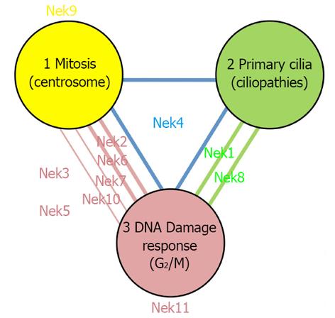
Figure 5 Functional overlap in the human Nek kinase family: seven of eleven Neks participate in two and one Nek in all three of the main core functions of the Nek family (centrosome-related mitosis, primary cilia and DNA damage response).
The three corners of the triangle represent each a key concept function for the Nek family, e.g., Nek9 and 11 sole involvement in mitosis[66,67] and DDR[73] respectively, has been well documented. The Nek names and bold lines represent cases where accumulated experimental evidence strongly suggests a regulatory role for that Nek in that context or in both of the contexts the line connects: Nek1[2,22,23]; Nek2[123]; Nek4[9,53] (Basei et al unpublished); Nek6[3]; Nek7[67]; Nek8[8,10]; Nek10[74]. The thinner lines represent our own group’s preliminary or unpublished interaction data (both from yeast two-hybrid system and immunoprecipitation coupled to mass spectrometry analysis data), suggestive of a participation of that Nek in both connected functions (Nek7: Souza et al, unpublished).
- Citation: Meirelles GV, Perez AM, de Souza EE, Basei FL, Papa PF, Melo Hanchuk TD, Cardoso VB, Kobarg J. “Stop Ne(c)king around”: How interactomics contributes to functionally characterize Nek family kinases. World J Biol Chem 2014; 5(2): 141-160
- URL: https://www.wjgnet.com/1949-8454/full/v5/i2/141.htm
- DOI: https://dx.doi.org/10.4331/wjbc.v5.i2.141


